Custom Programs
Curriculum
Contact
Register

i2 Learning

Custom Programs
Curriculum
Contact
Register
Heart Dissection

Heart Dissection

Students prepare to dissect a sheep heart
Heart Dissection

Heart Dissection

Students prepare to dissect a sheep heart

Heart Dissection

Heart Dissection

Students dissect the sheep heart

Heart Dissection

Heart Dissection

Students dissect the sheep heart together
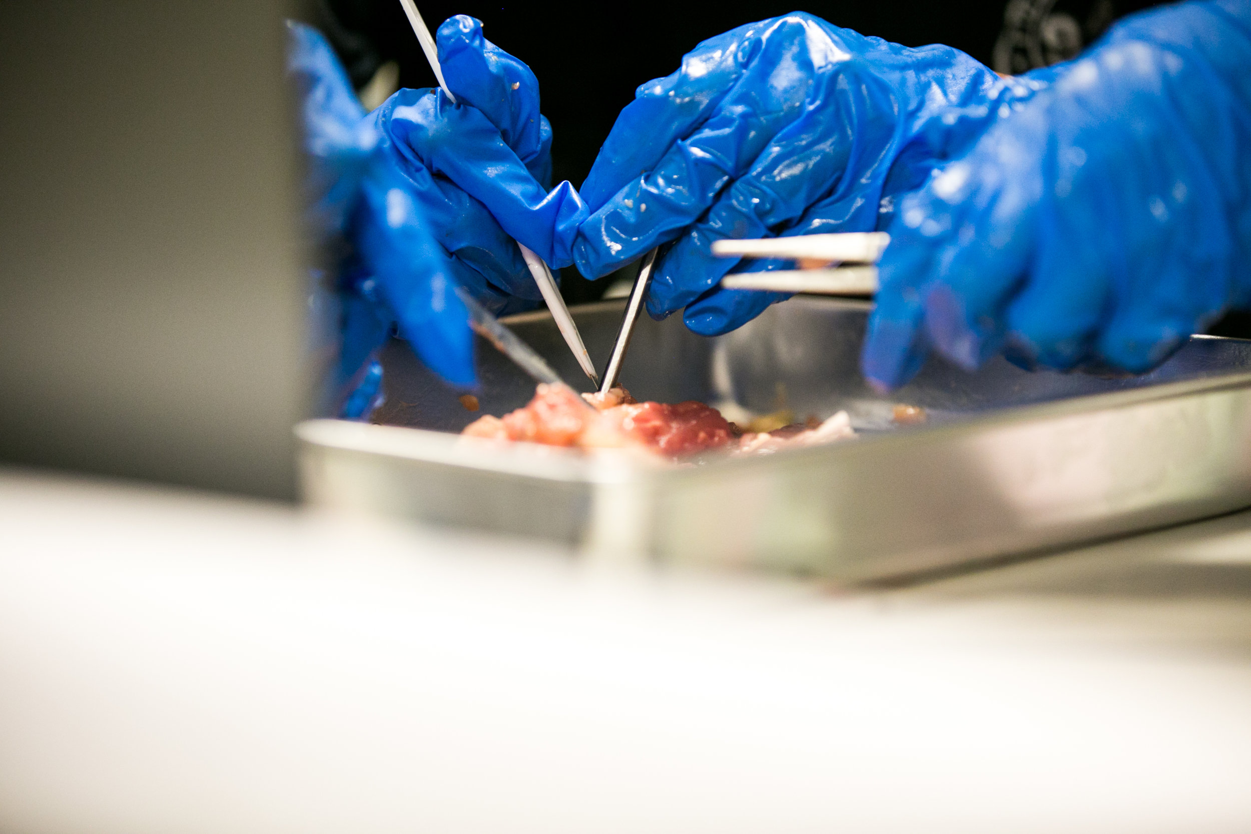
Heart Dissection

Heart Dissection

Students work together to dissect the sheep heart
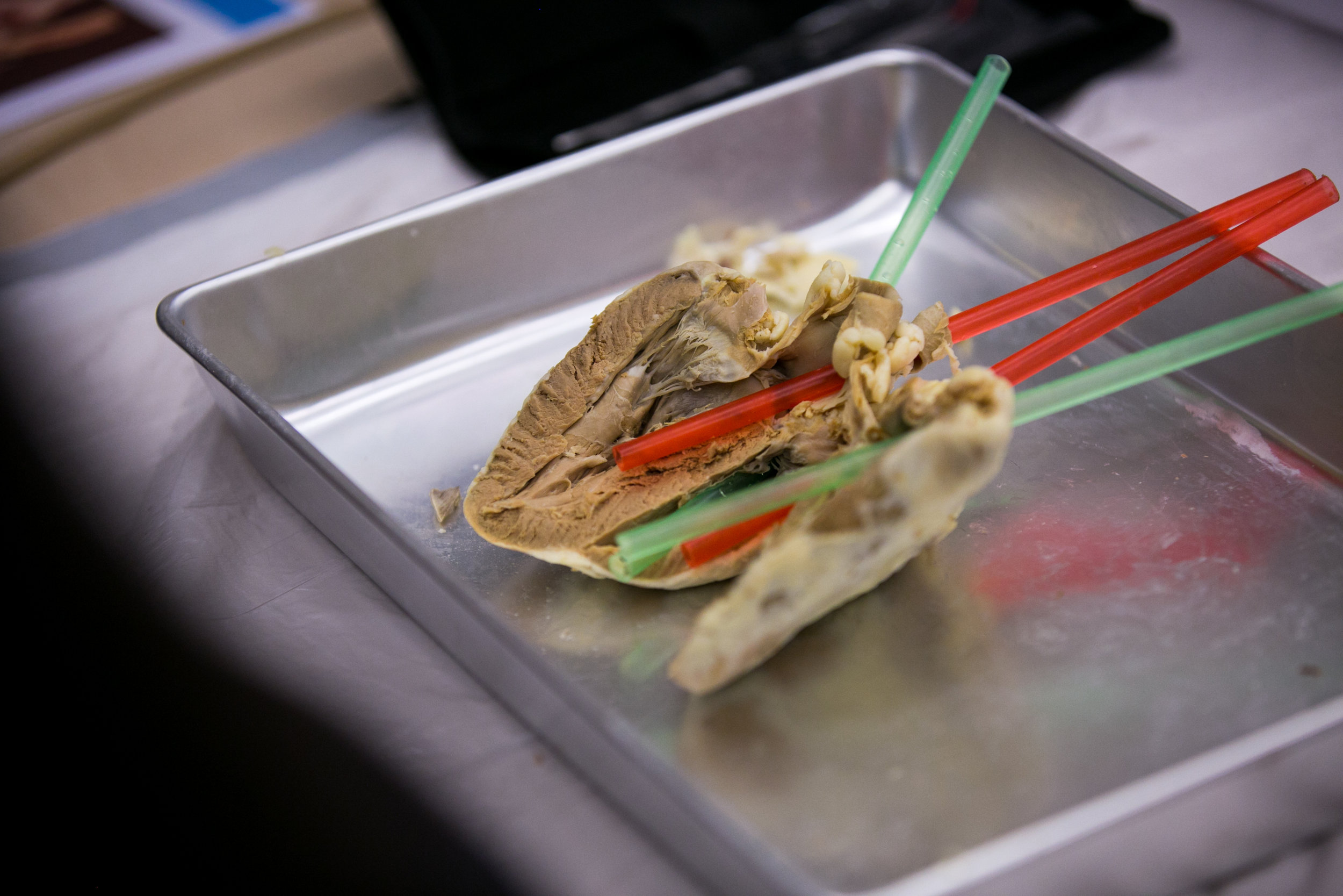
Heart Dissection

Heart Dissection

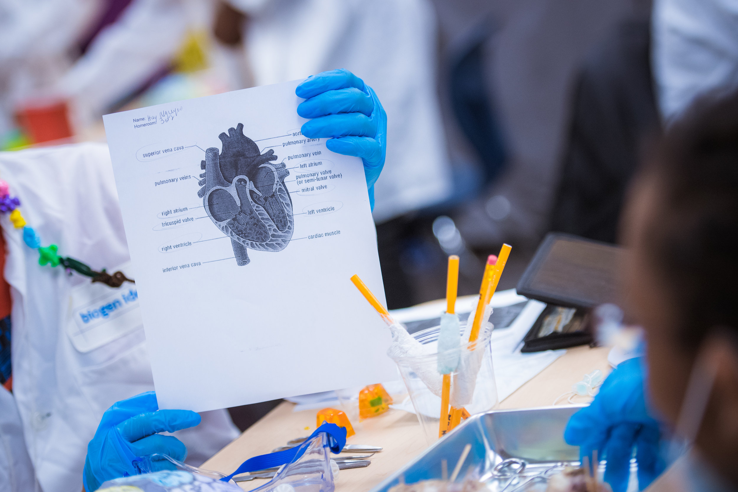
Heart with Blood Vessel Labels

Heart with Blood Vessel Labels

Heart with labels aorta, superior vena cava, pulmonary artery, and pulmonary vein.
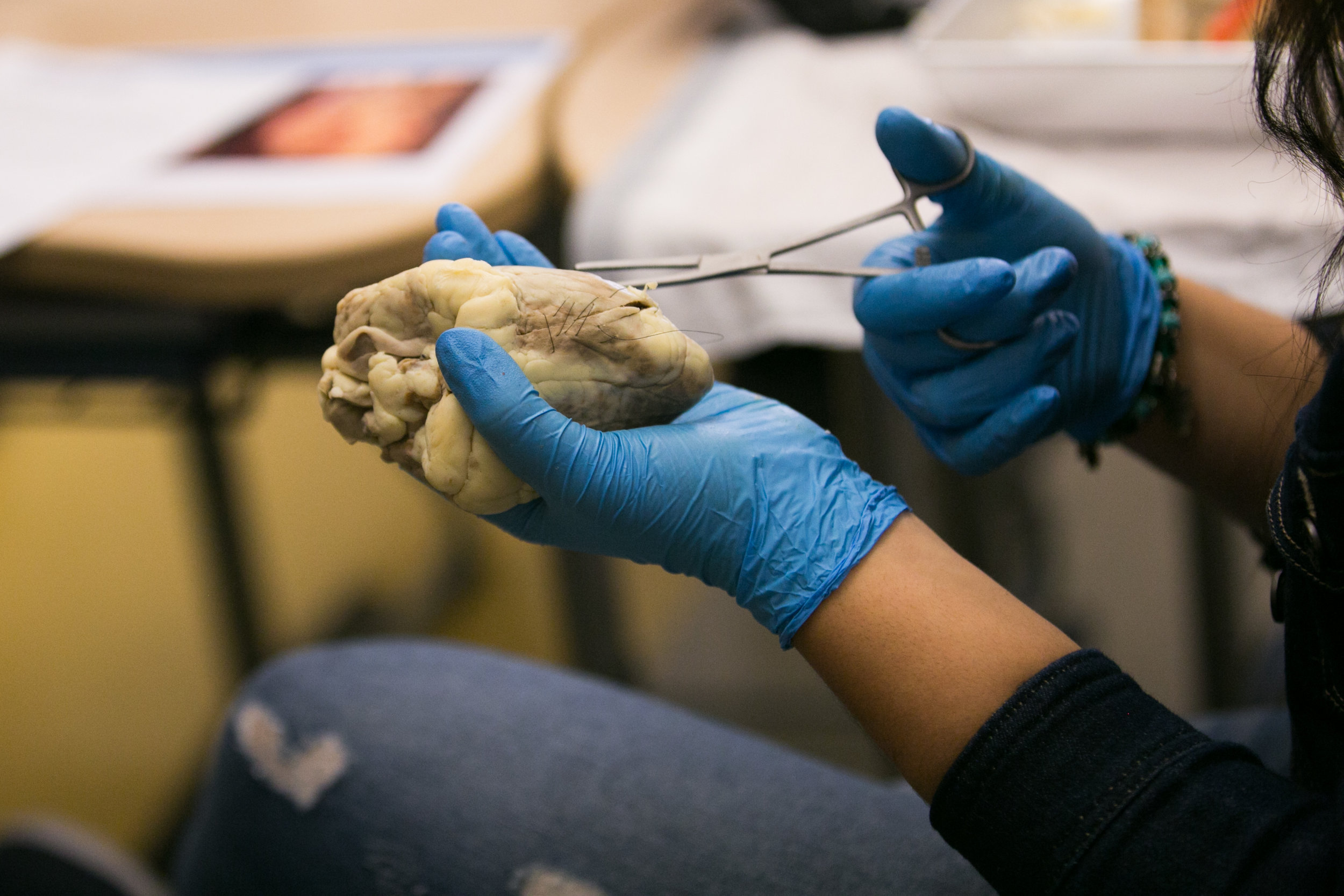
Heart Dissection

Heart Dissection

Student dissects a sheep heart
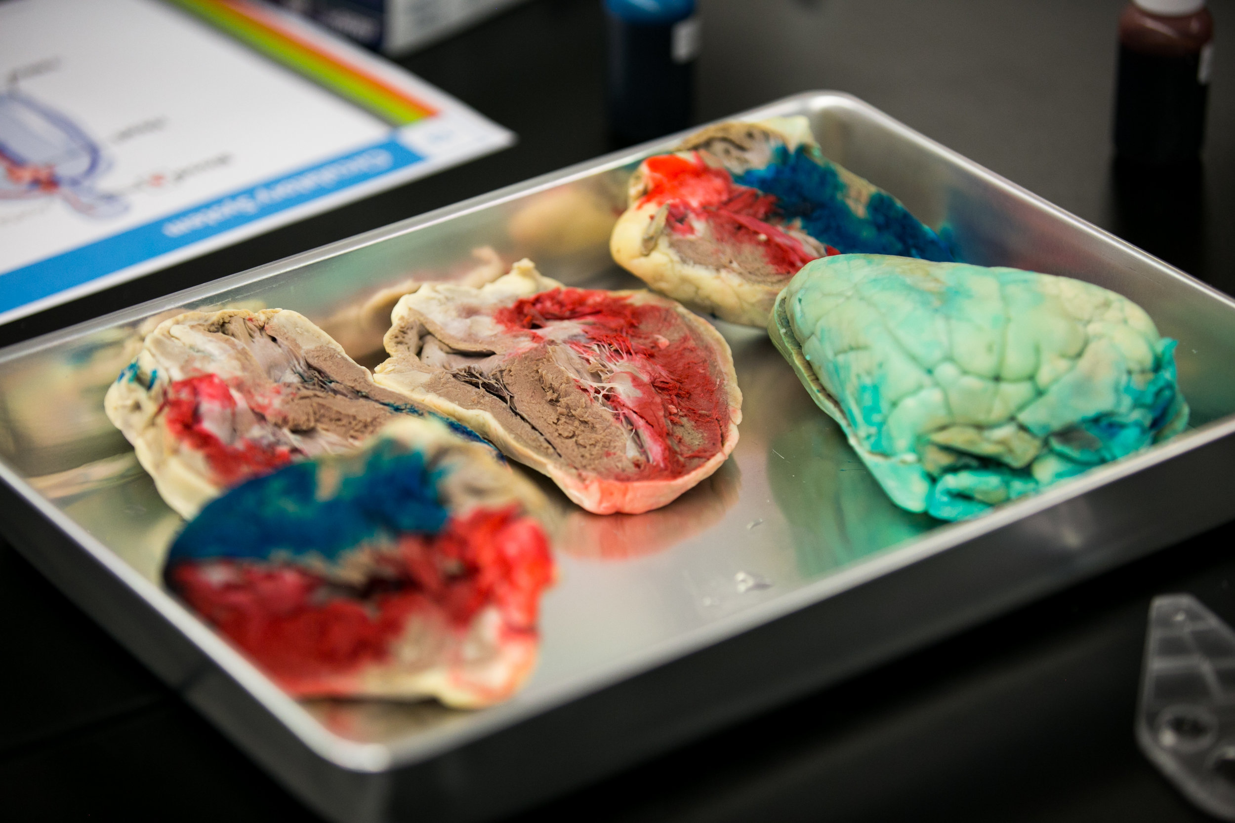
Heart Dissection

Heart Dissection

Heart Dissection

Heart Dissection

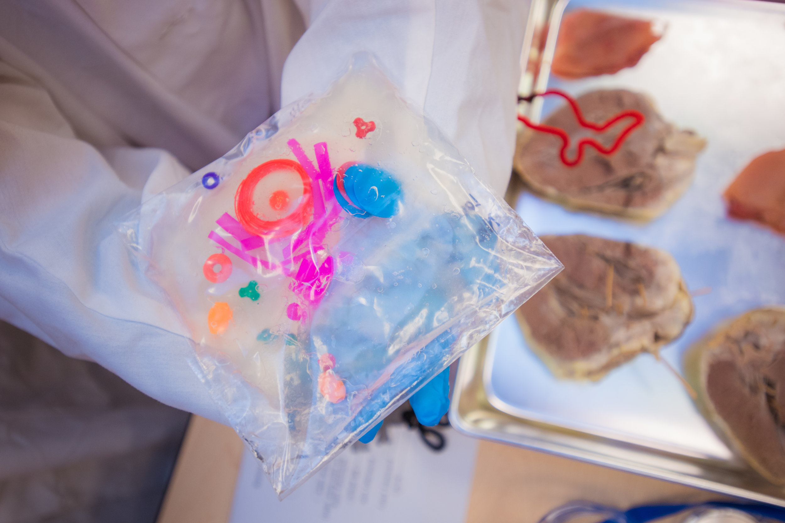
Student shows the dissected sheep heart with different colored dyes used to show the different parts of the heart

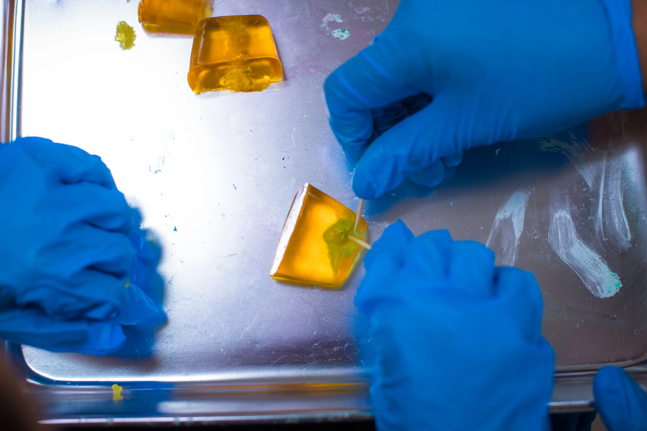
Systems Working Together: Biopsy Simulation

Systems Working Together: Biopsy Simulation

The first incision of a biopsy

Systems Working Together

Systems Working Together

Students conduct a model biopsy and lumpectomy
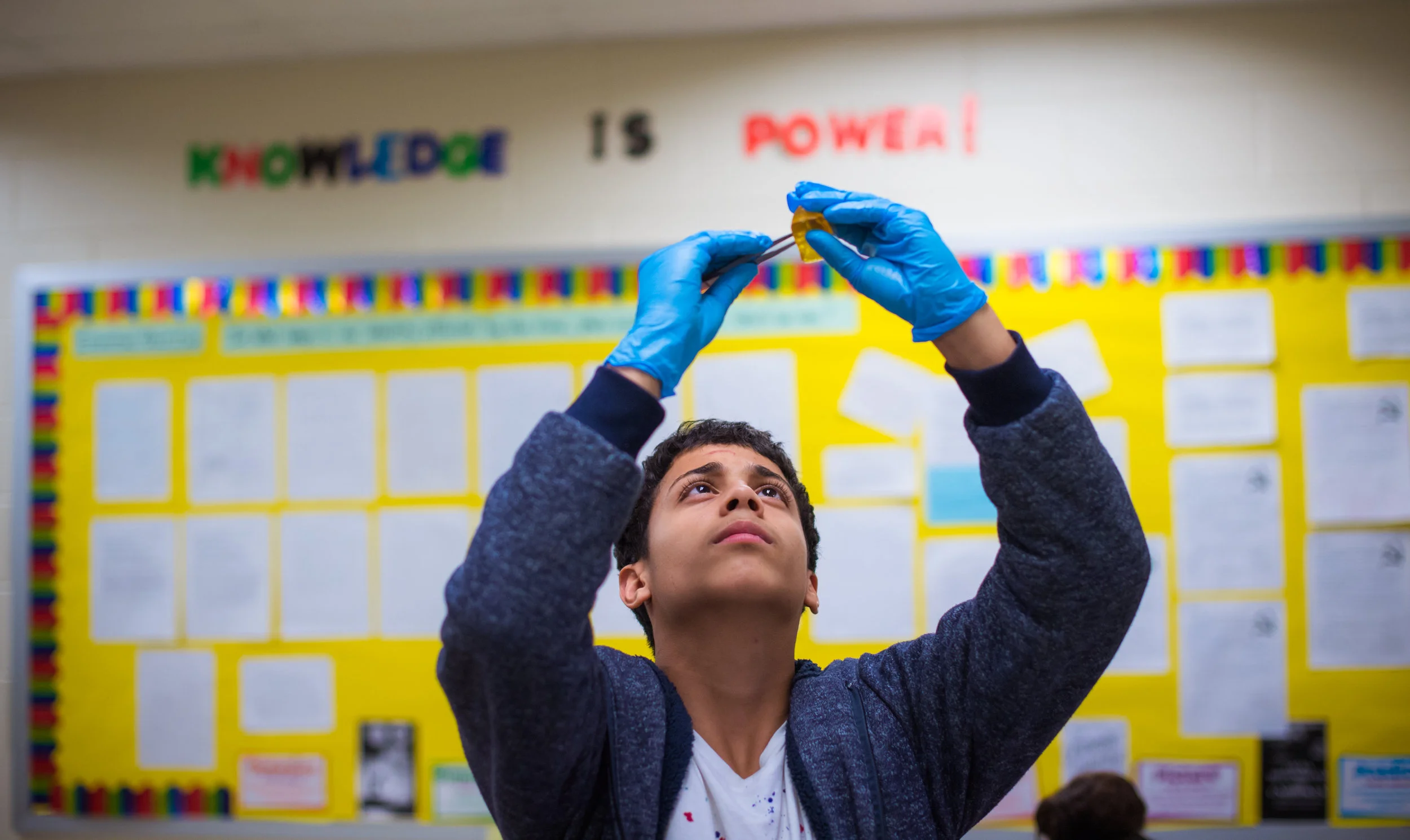
Systems Working Together

Systems Working Together

A student uses tweezers to perform a biopsy simulation.
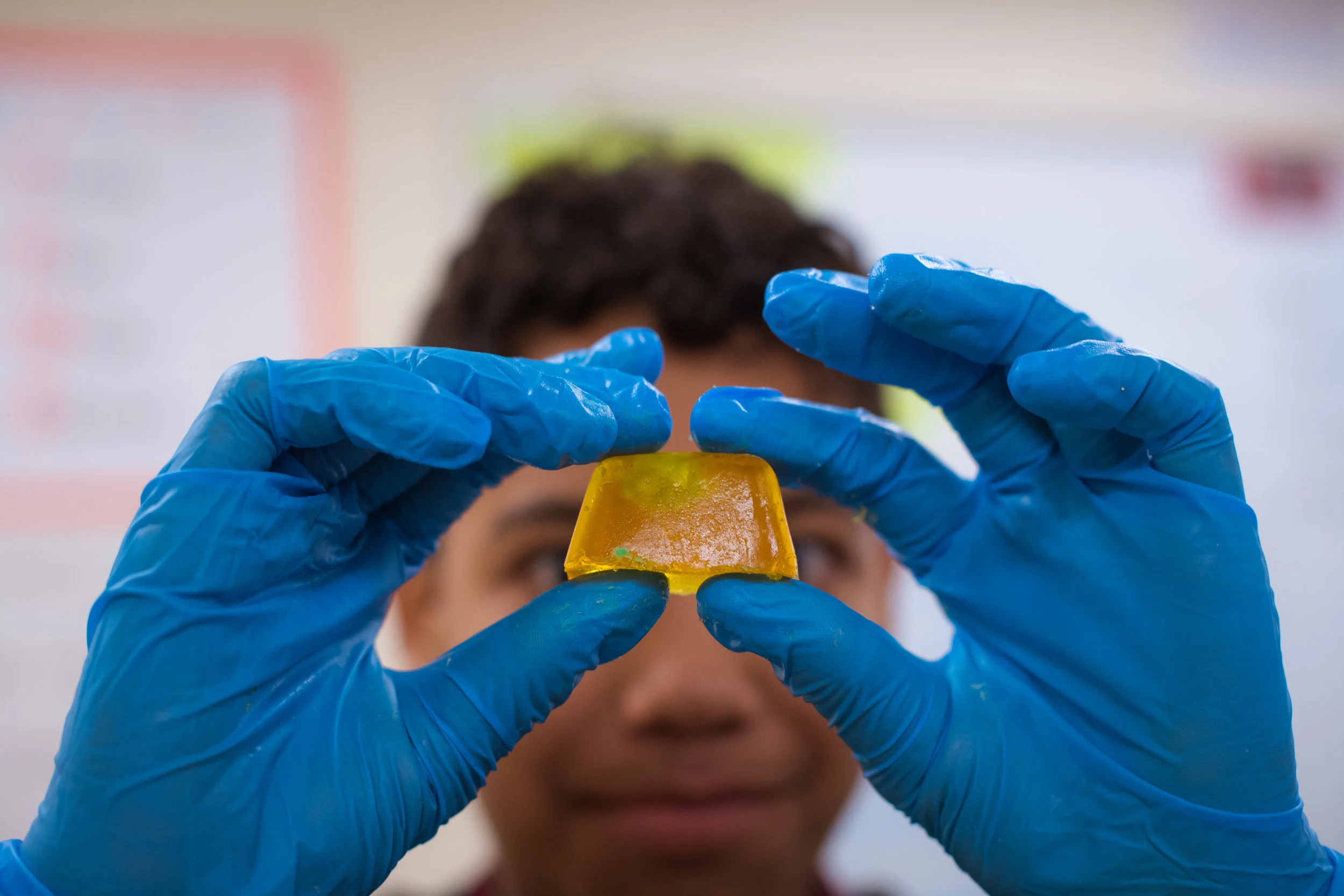
Systems Working Together

Systems Working Together

Student prepares to remove a piece of the play doh from the gelatin cube to simulate a biopsy
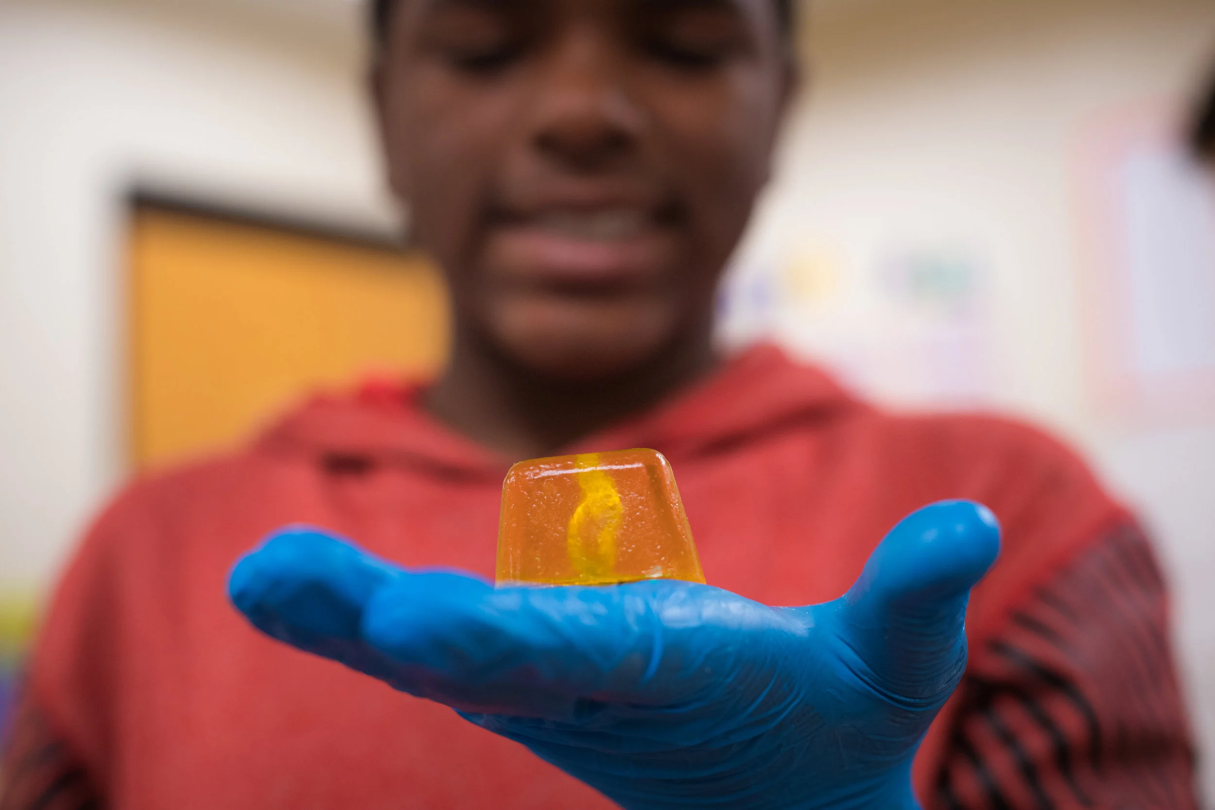
Systems Working Together

Systems Working Together

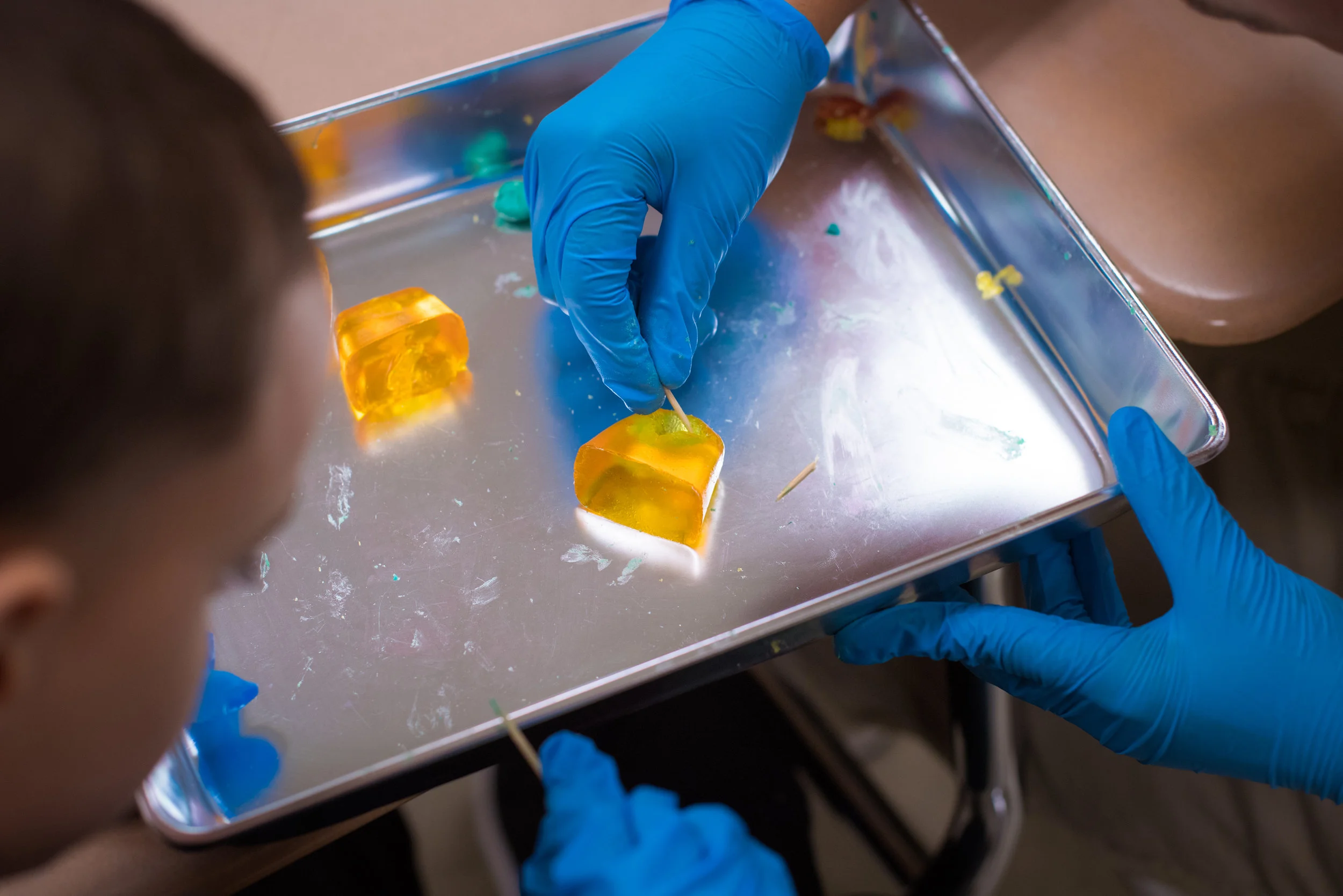
Student prepares to perform a lumpectomy
Systems Working Together

Systems Working Together

Students perform a biopsy

How to Suture

How to Suture

Student shows how to suture

Model Neurons

Model Neurons

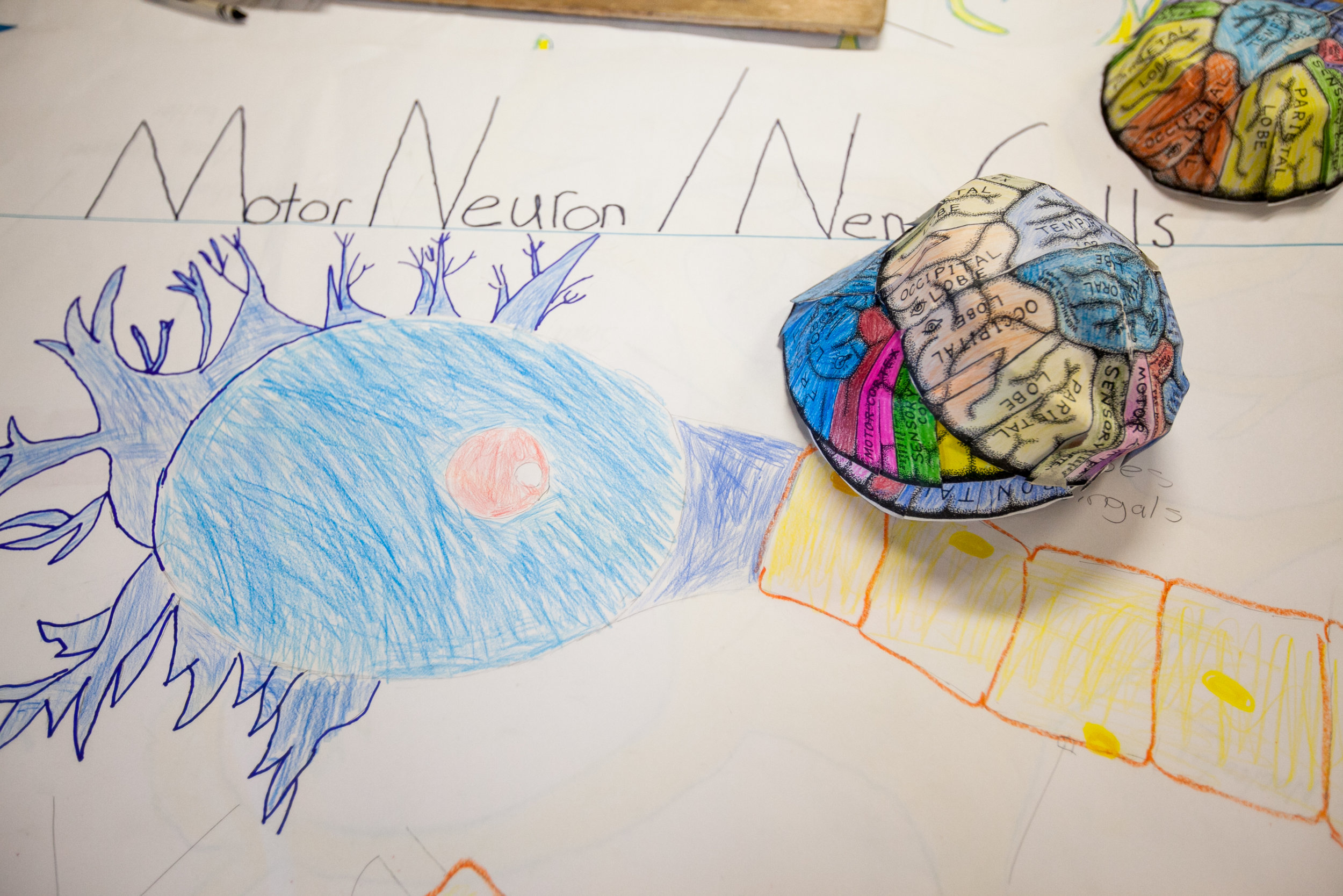
A completed model neuron

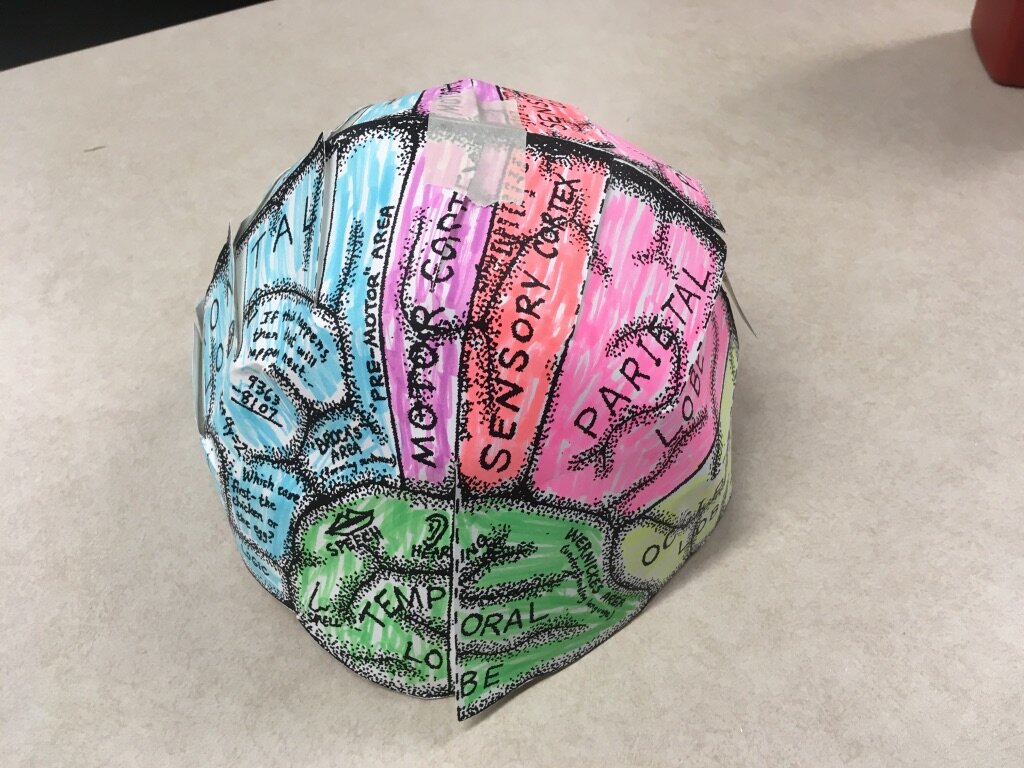
Brain Hats

Brain Hats

Students create brain hats that show the different parts of the brain
Brain Hats
Brain Hats

Brain Hats

Brain Hats

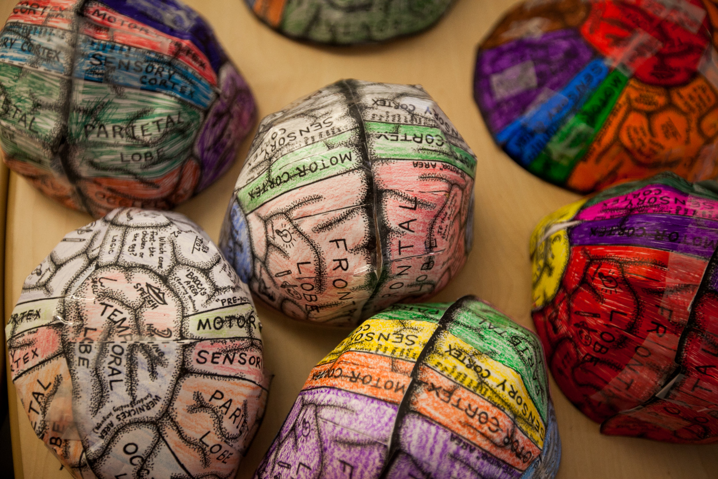
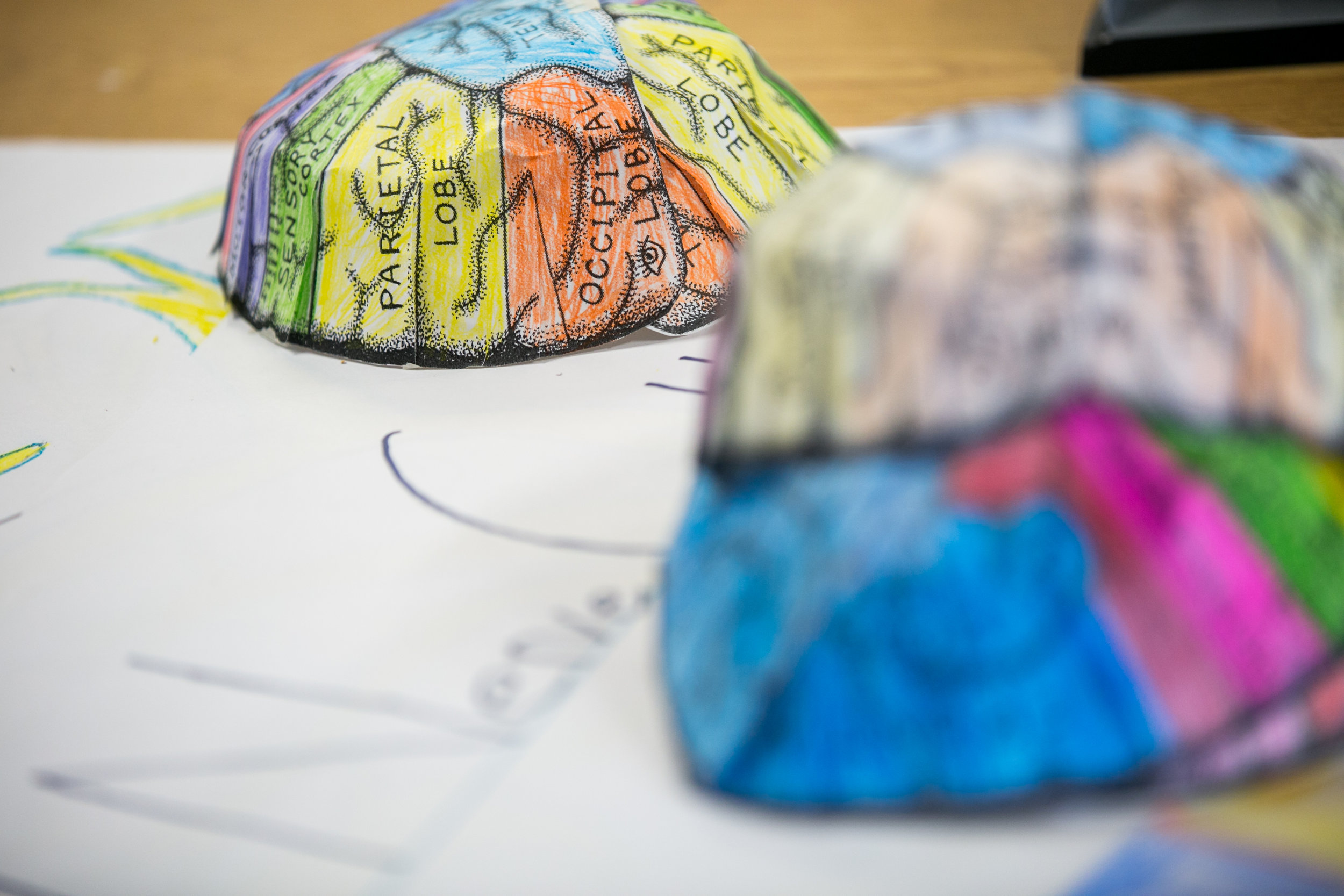
Completed brain hats with the different structures colored in

Brain Hats

Brain Hats

Student wears their completed brain hat
Brain Hats

Brain Hats

Brain Hats

Brain Hats

Students wear their completed brain hats during other activites
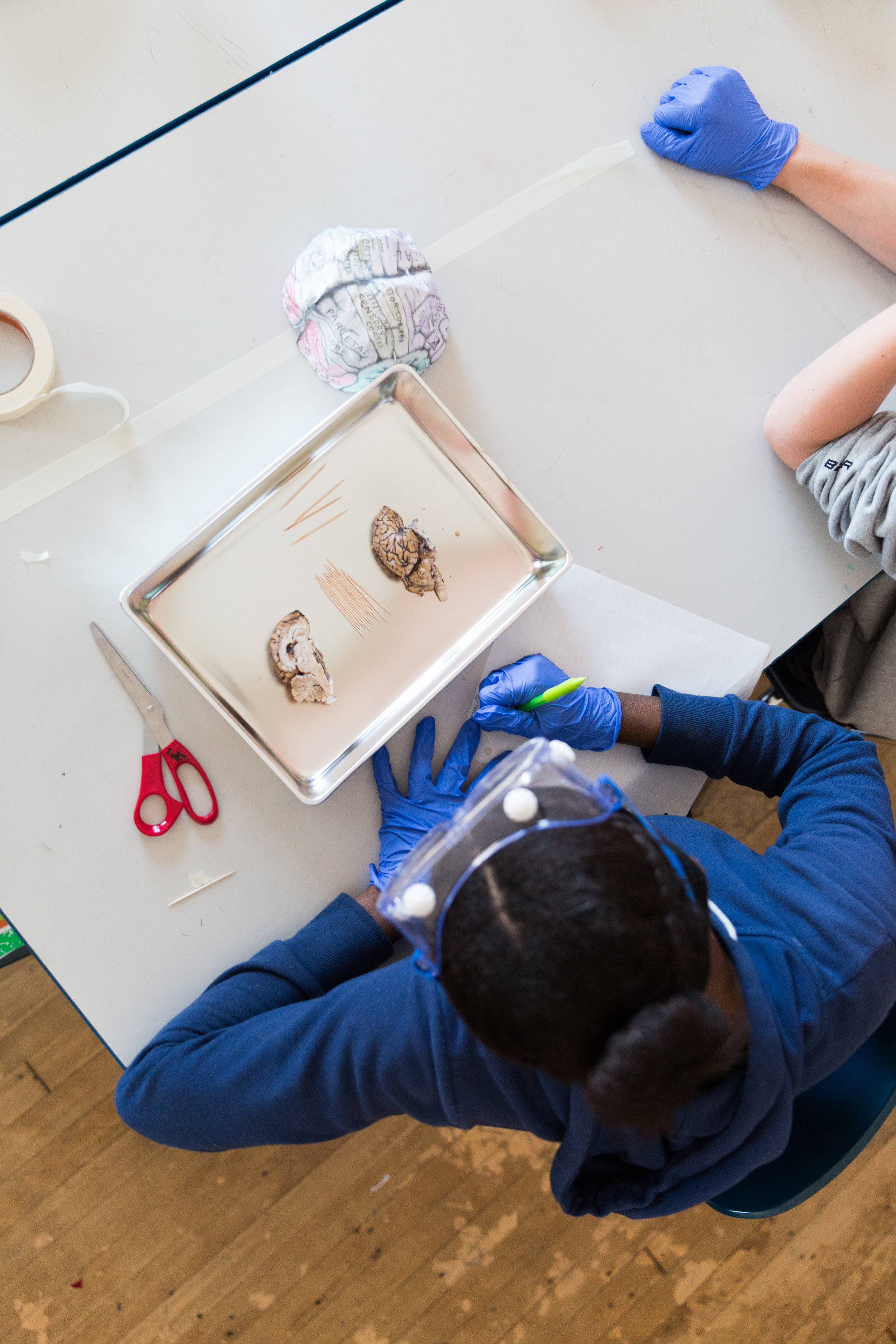
Sheep Brain Dissection

Sheep Brain Dissection

Students review the parts of the brain in preparation for dissection

Tools for the Sheep Brain Dissection

Tools for the Sheep Brain Dissection

Students dissect a sheep brain to observe the various lobes, grey and white matter, optic chasm and cerebellum among other anatomical structures. Students practice sagittal, axial and traverse cuts.

Sheep Brain Dissection

Sheep Brain Dissection

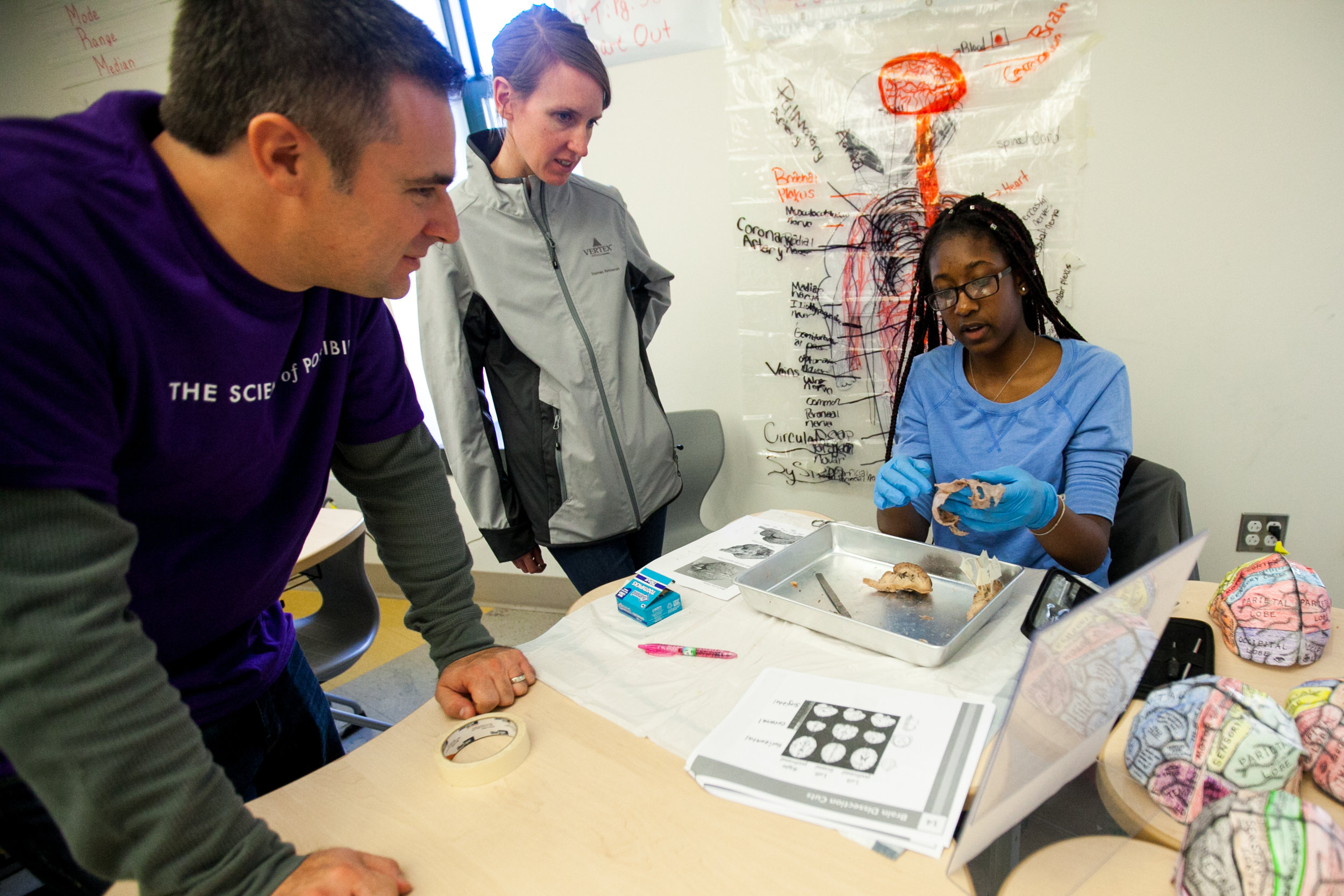
Teacher points out the different parts of the brain
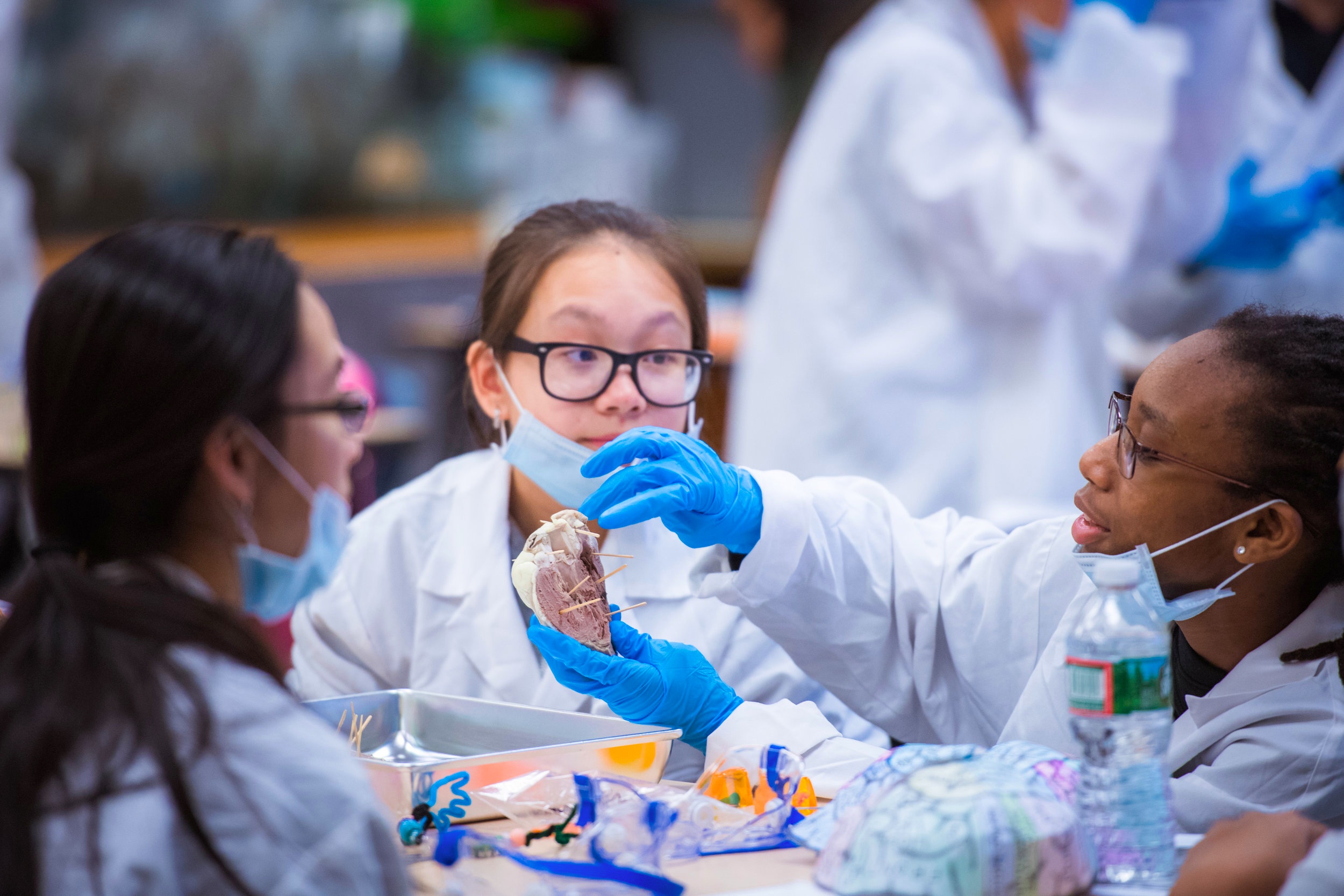
Sheep Brain Dissection

Sheep Brain Dissection

Students identify the different parts of the brain
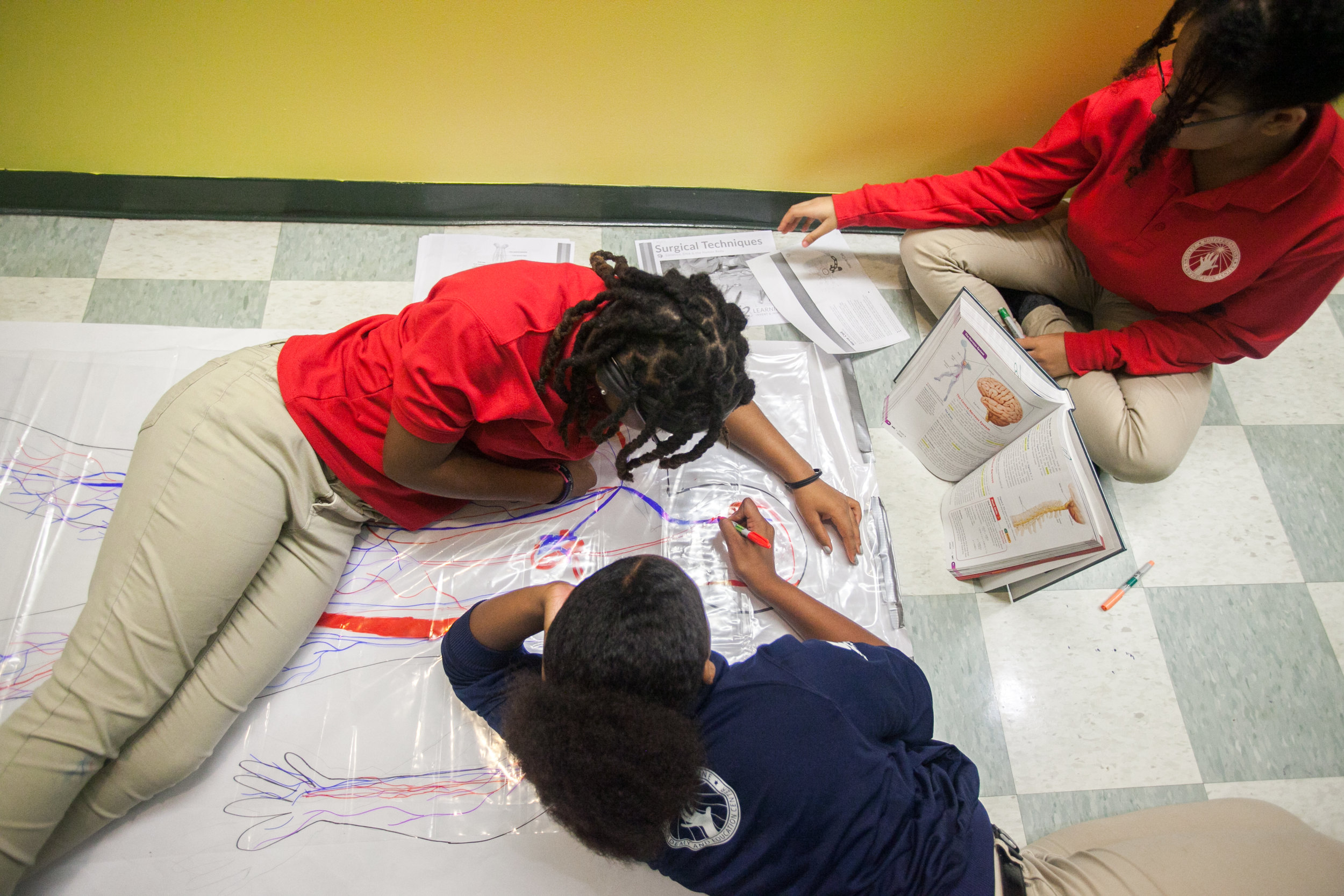
Model Patient

Model Patient

Students draw the different systems on their model patient
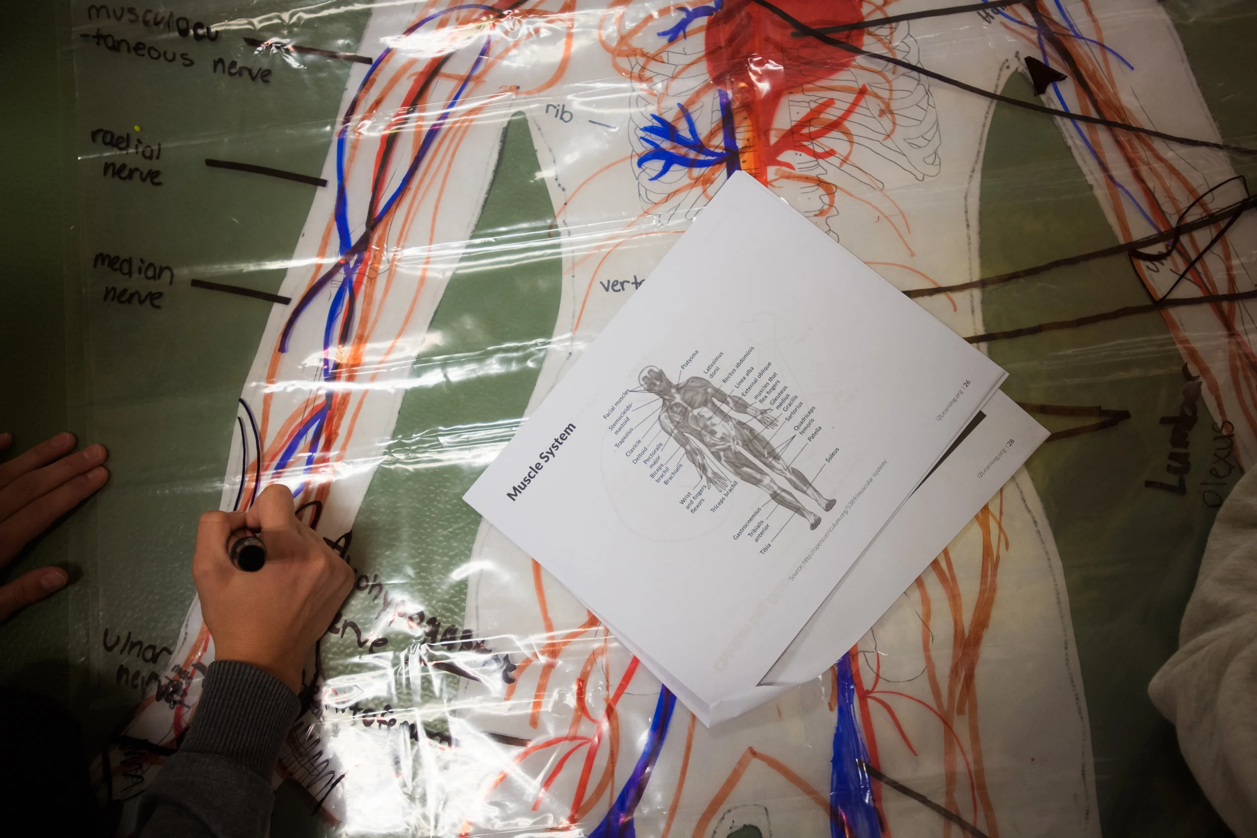
Model Patient

Model Patient

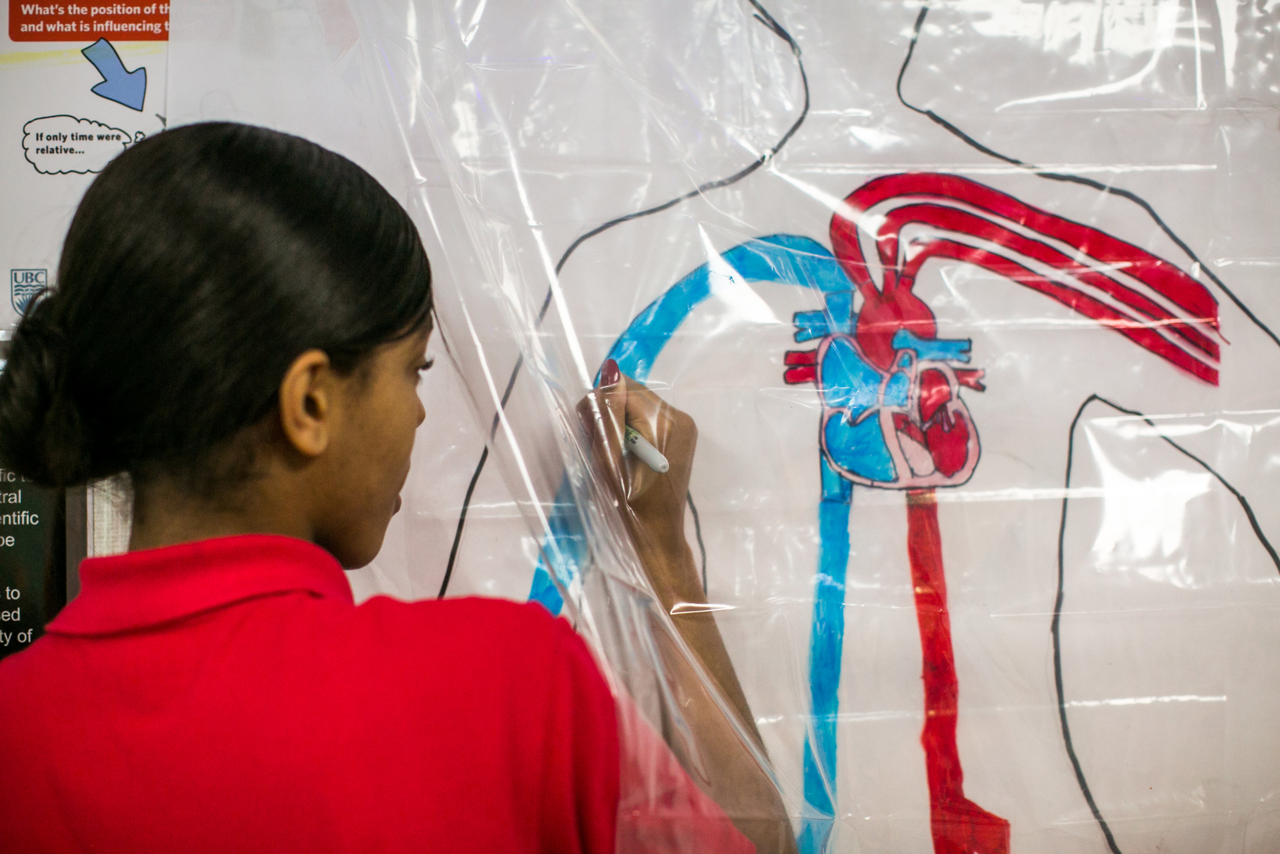
Students use shower curtains to draw the different systems of the body
Model Patient

Model Patient

Student draws the systems of the model patient

Model Patient

Model Patient

Students are presented with a hypothetical patient who has endured a traumatic accident and are called to diagnose and treat their patient based on the knowledge and experience they have obtained.
Model Patient

Model Patient

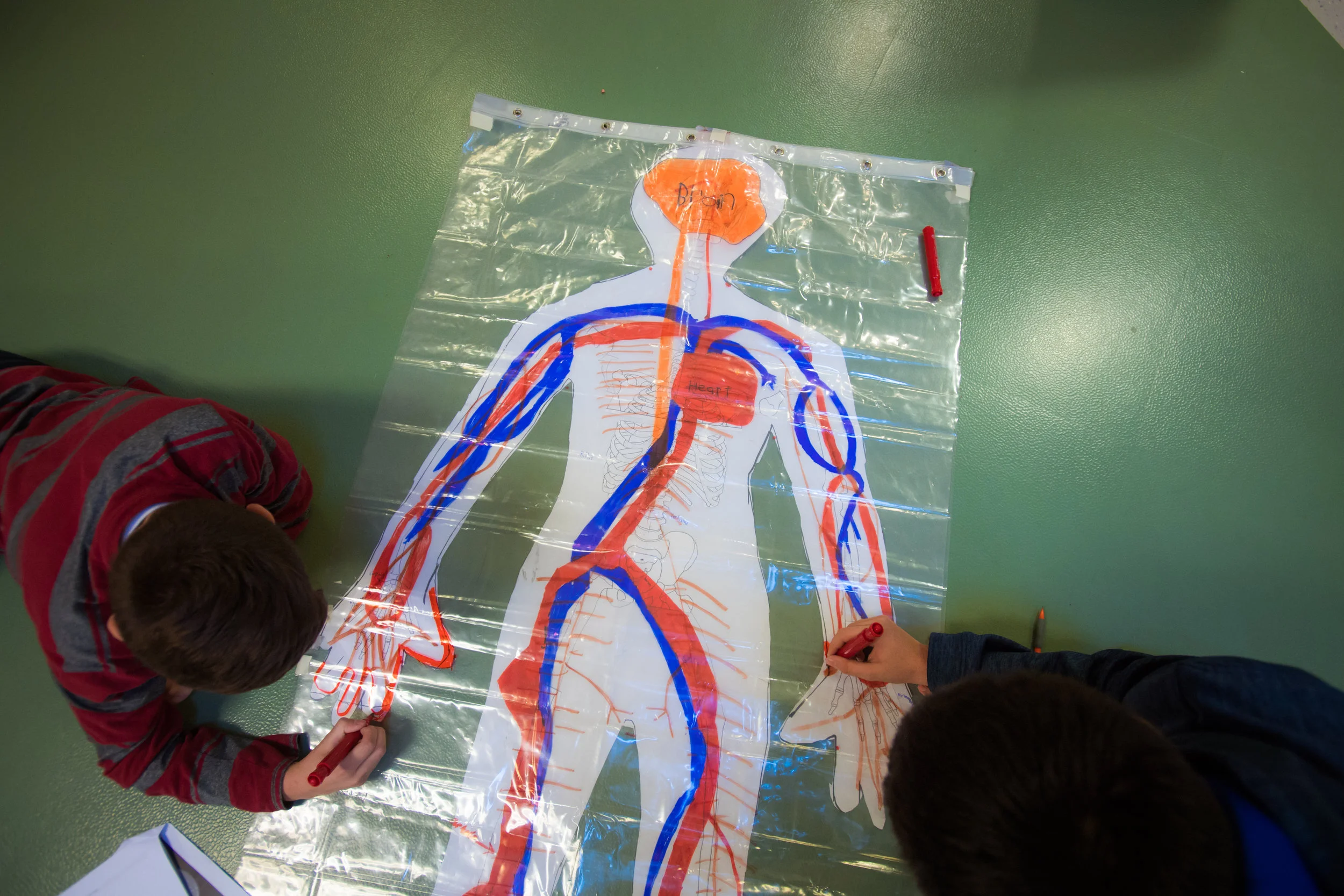
Students create a diagram using shower curtains to show how the body systems are related
Model Patient

Model Patient

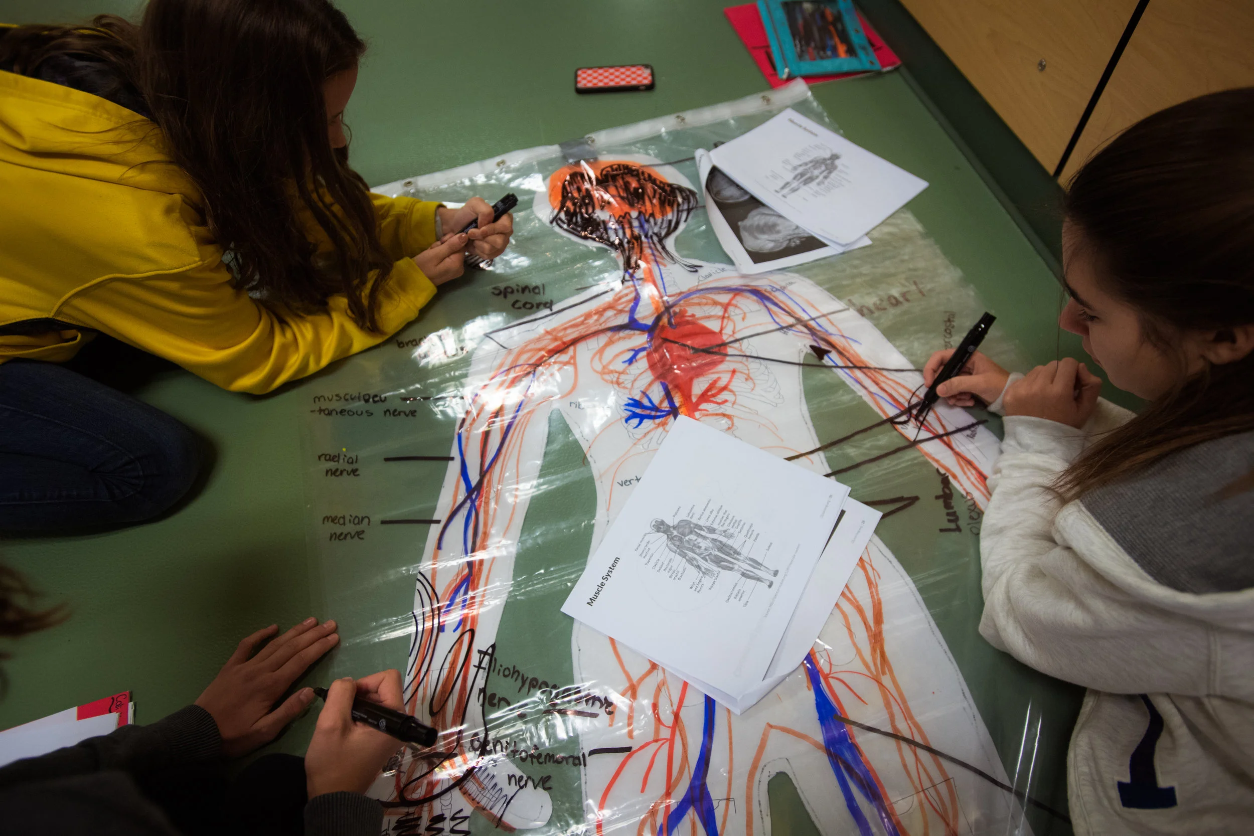
Students draw the systems on their model patient

Model Patient

Model Patient

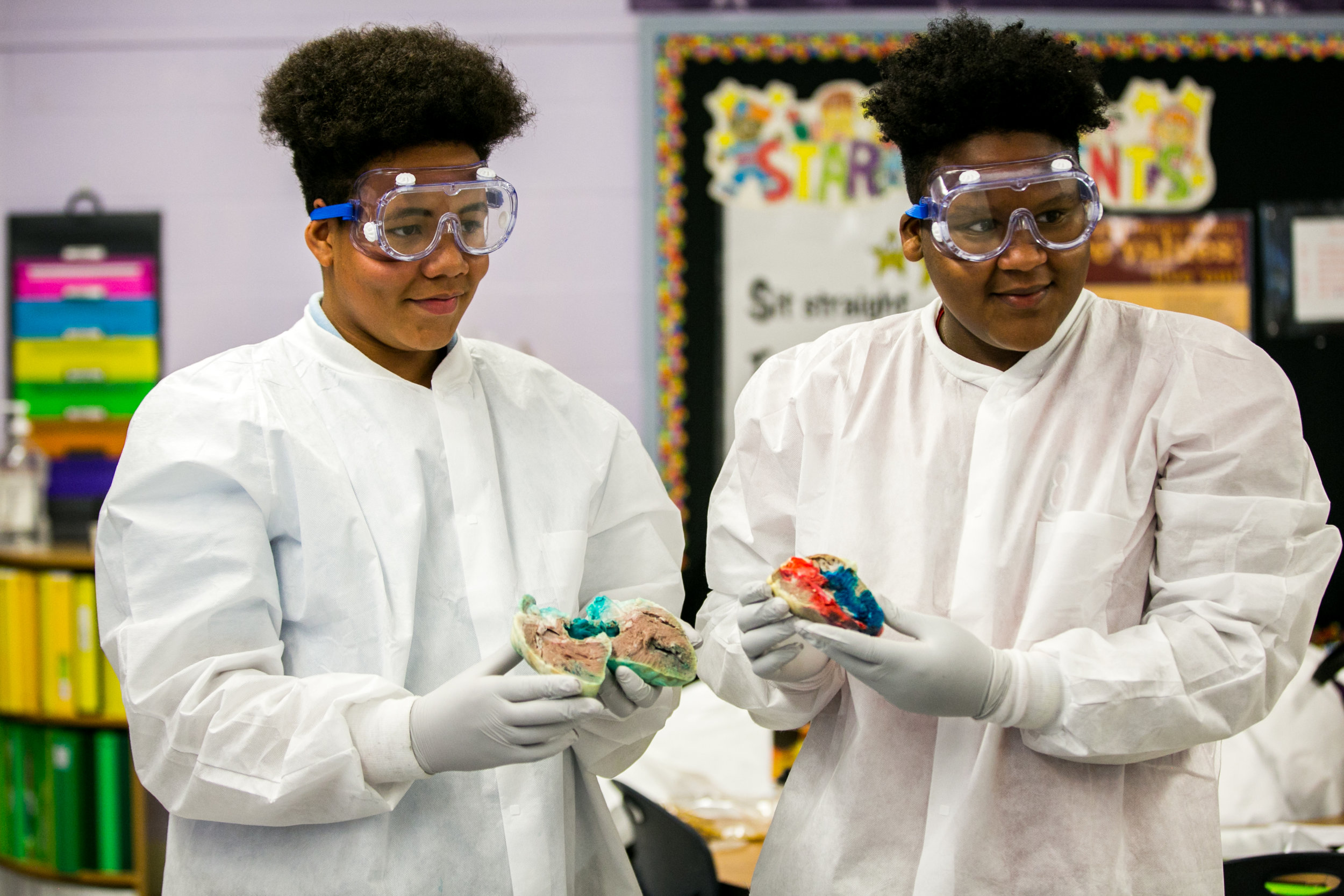
Completed model patient
Showcase

Showcase

Students show and explain the different parts of their dissected heart for the Showcase visitors.

Showcase

Showcase

Students use dye to show how the heart works.
Showcase

Showcase

Student shows guests the different parts of the brain
Showcase

Showcase

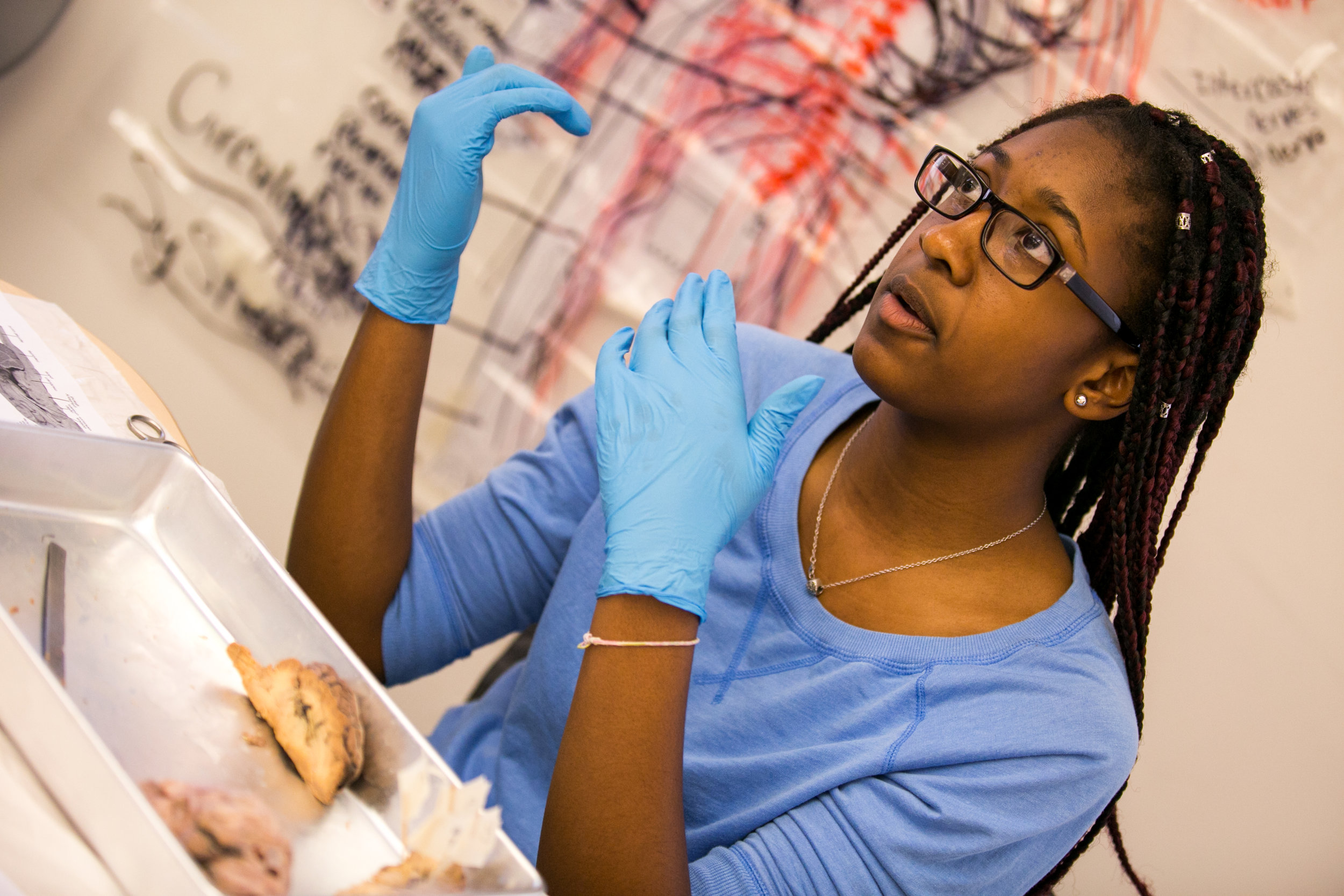
Student talks about the brain dissection during showcase
Showcase

Showcase

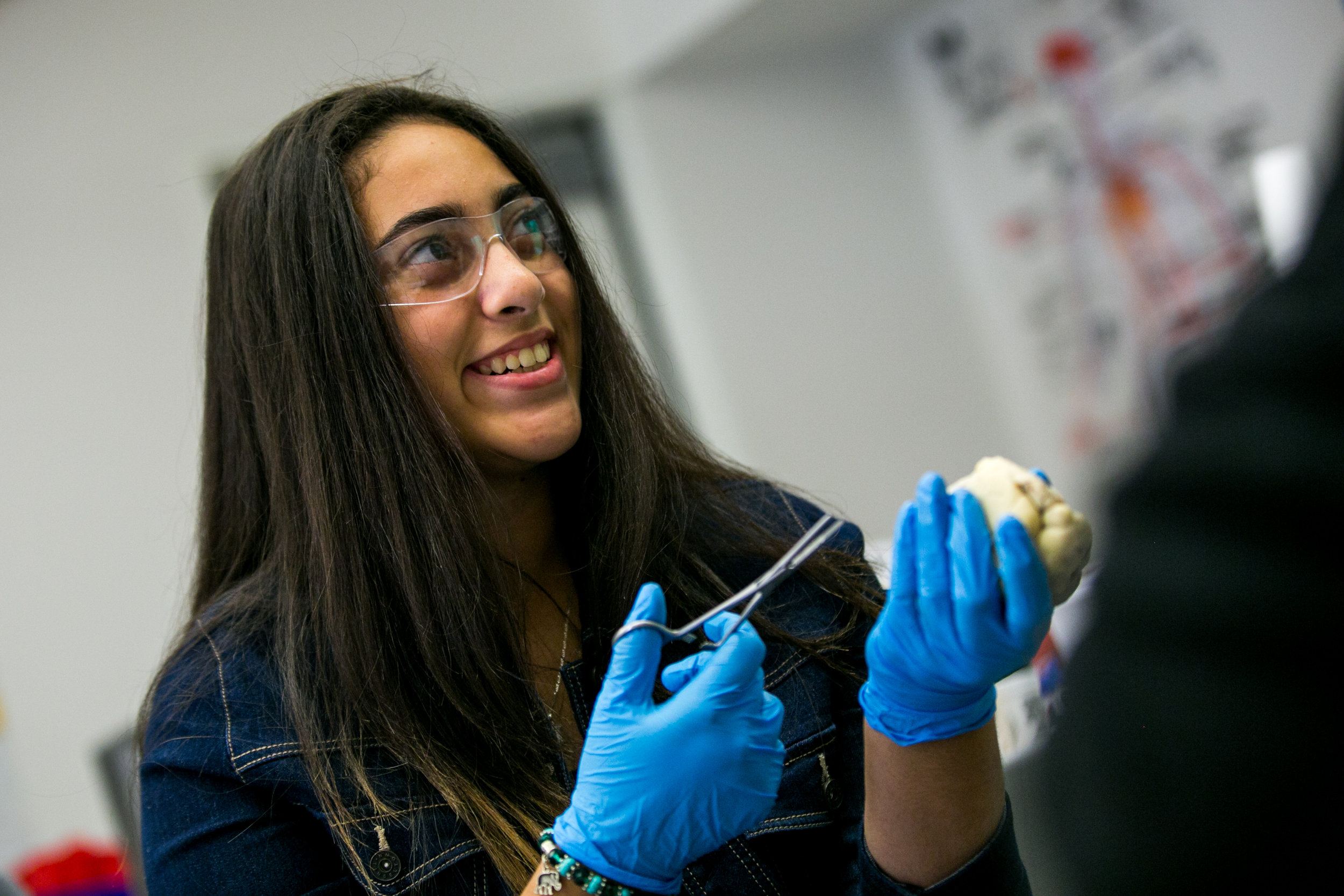
Student shows the sheep heart to a visitor during showcase
Showcase

Showcase

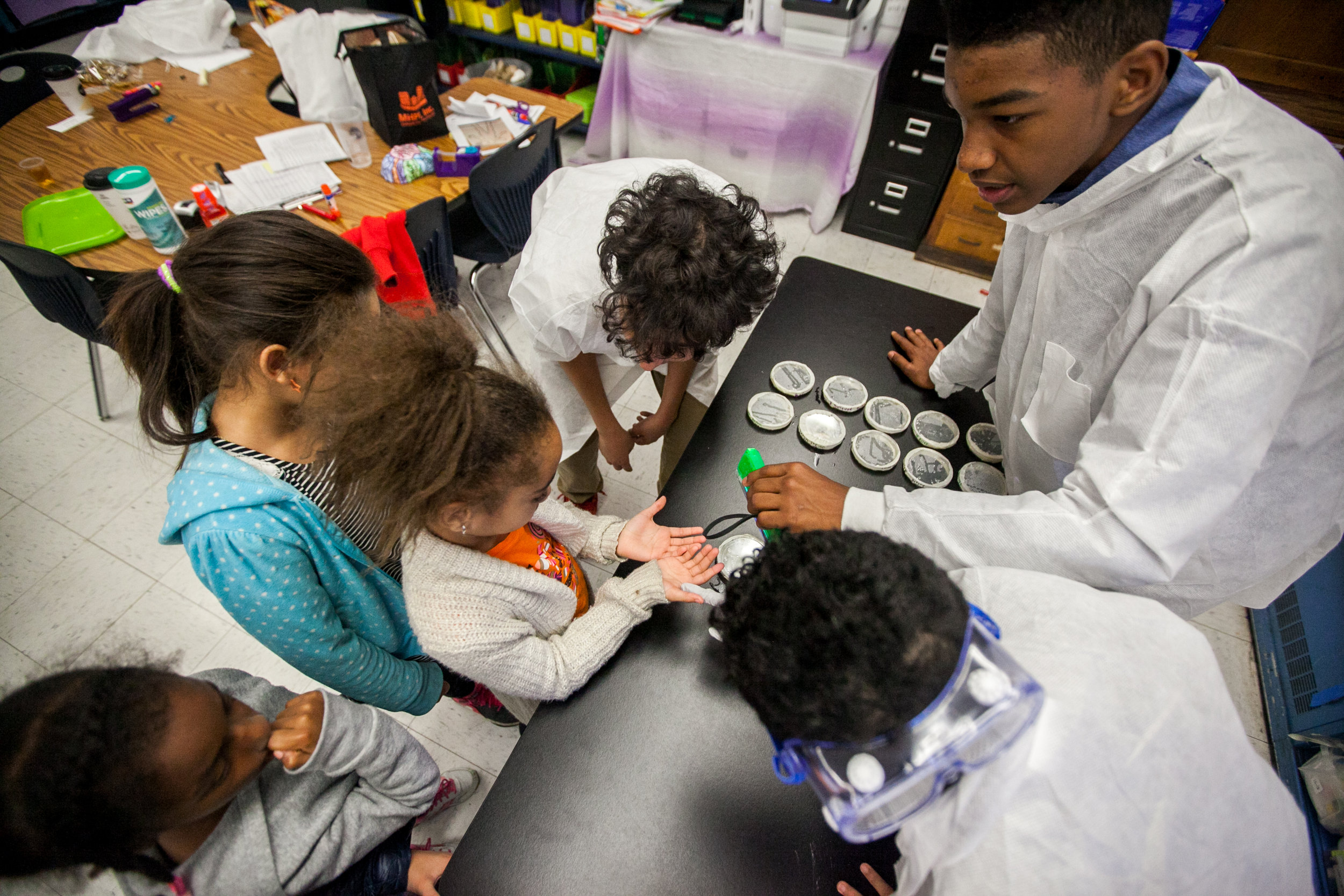
Student shows the younger students what they’ve learned this week
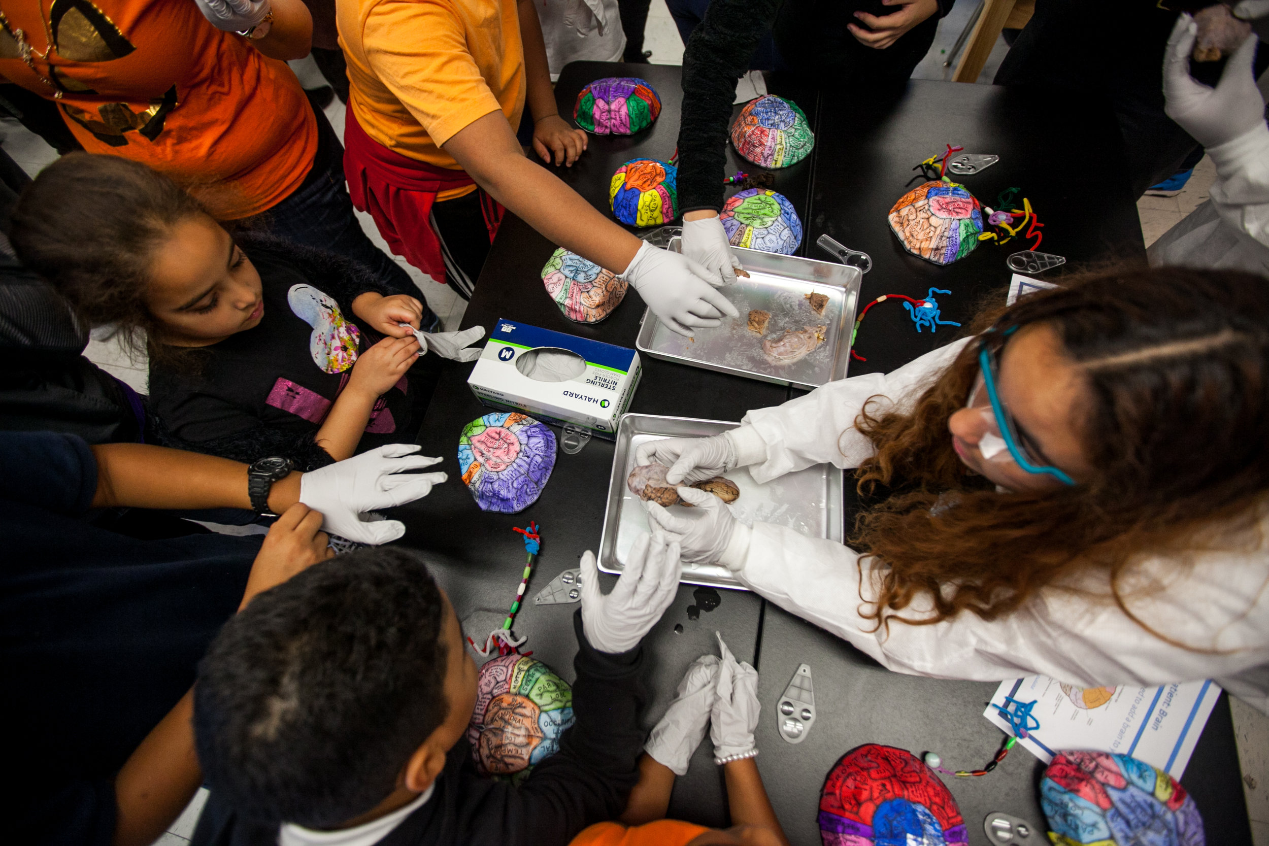
Showcase

Showcase

Student shows guests the different parts of the brain using the brain hats and the sheep brain

Showcase

Showcase

Student shows guest the brain hats and the dissected sheep brain during the showcase
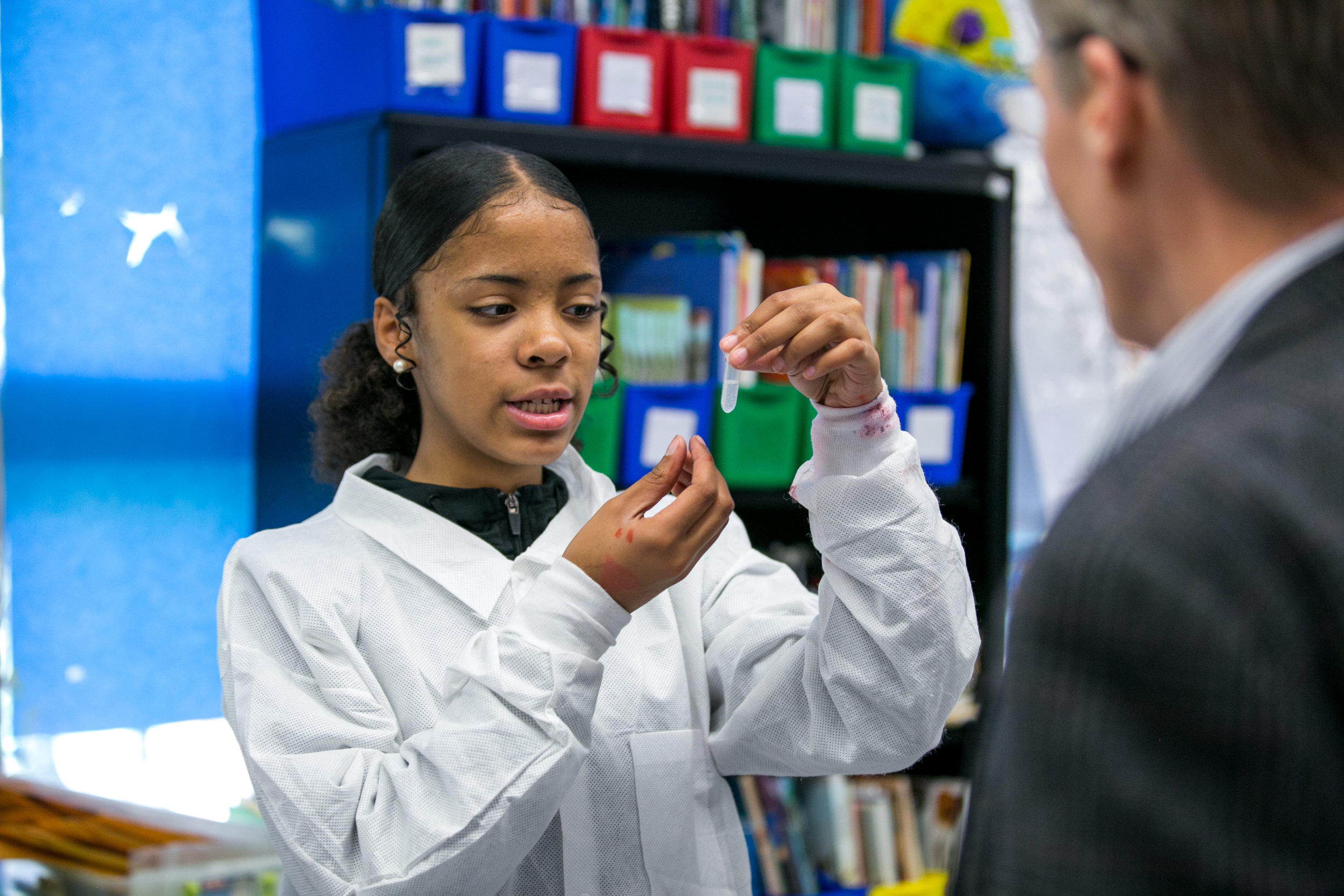
Showcase

Showcase

Student shows her DNA to visitor during the showcase
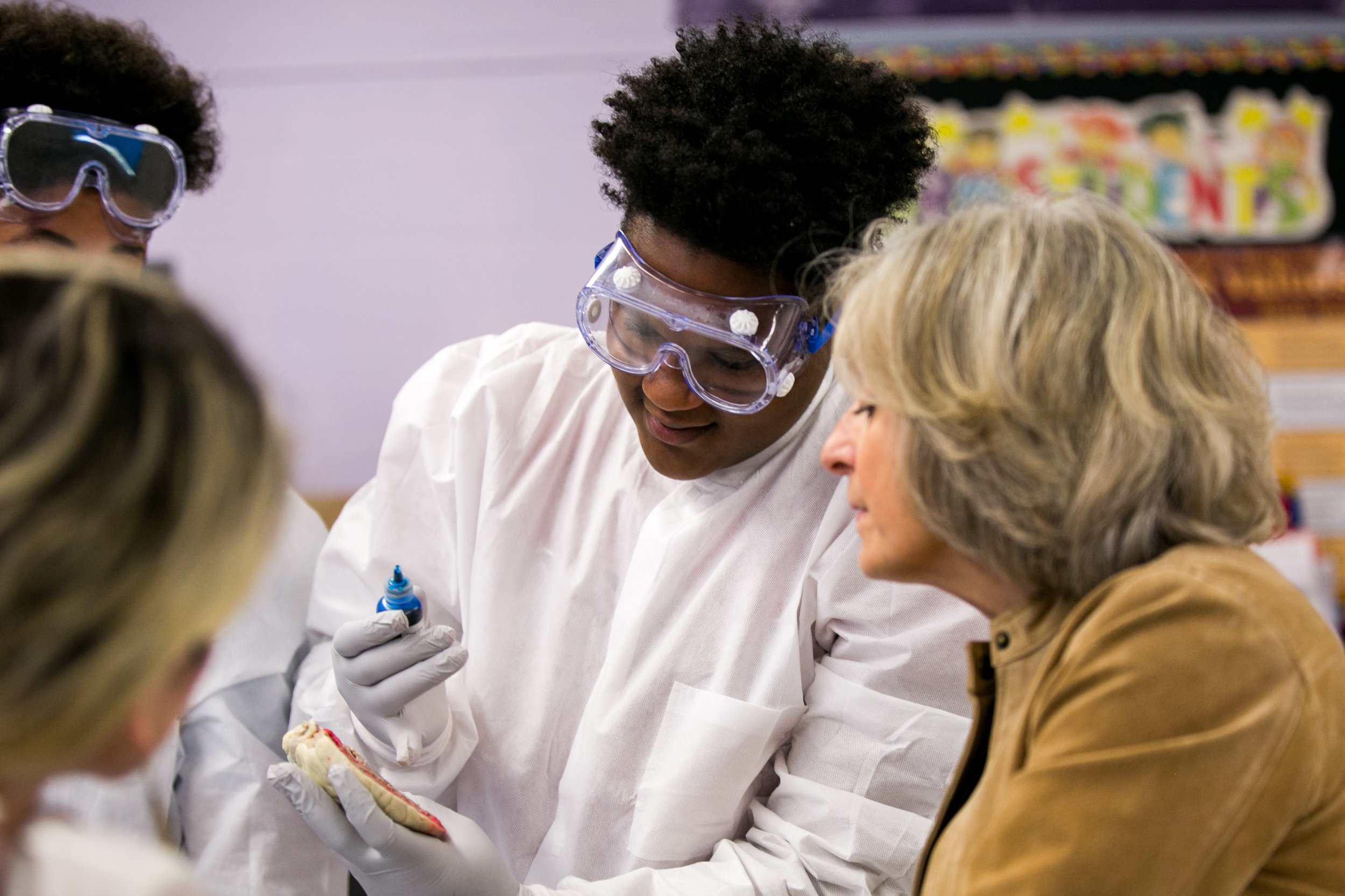
Showcase

Showcase

Student shows visitor how dyes are used to show the different parts of the heart
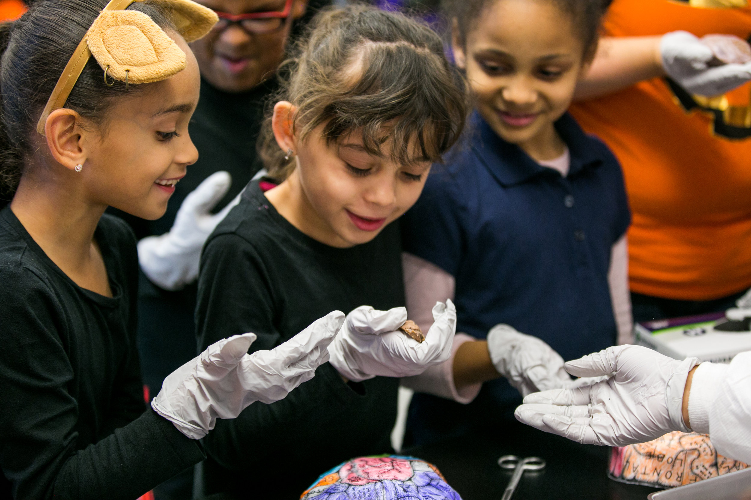
Showcase

Showcase

Showing the sheep brain that was dissected to younger students during the showcase

Grybus-i2-Hurley-HR-4357.jpg
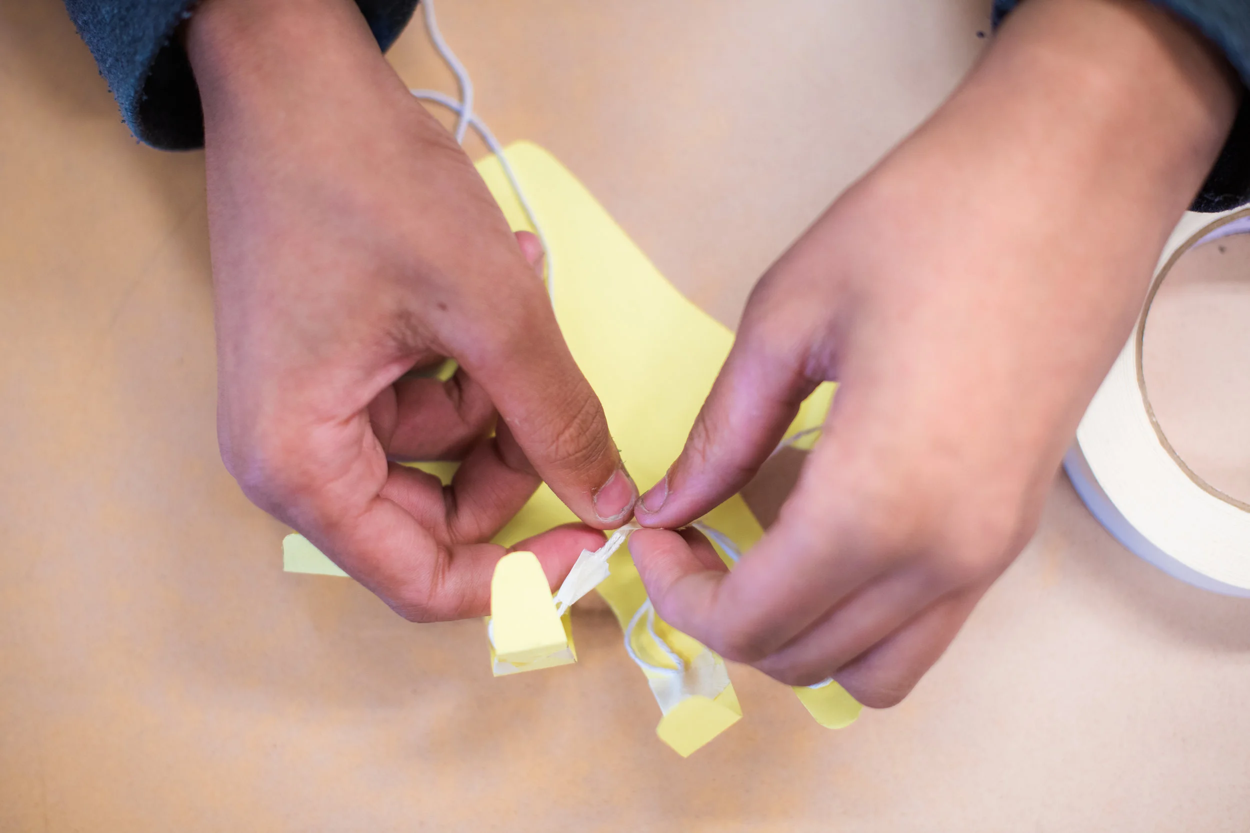
Creating the Model Hand

Creating the Model Hand

Tightening the string to create the movements of the model hand
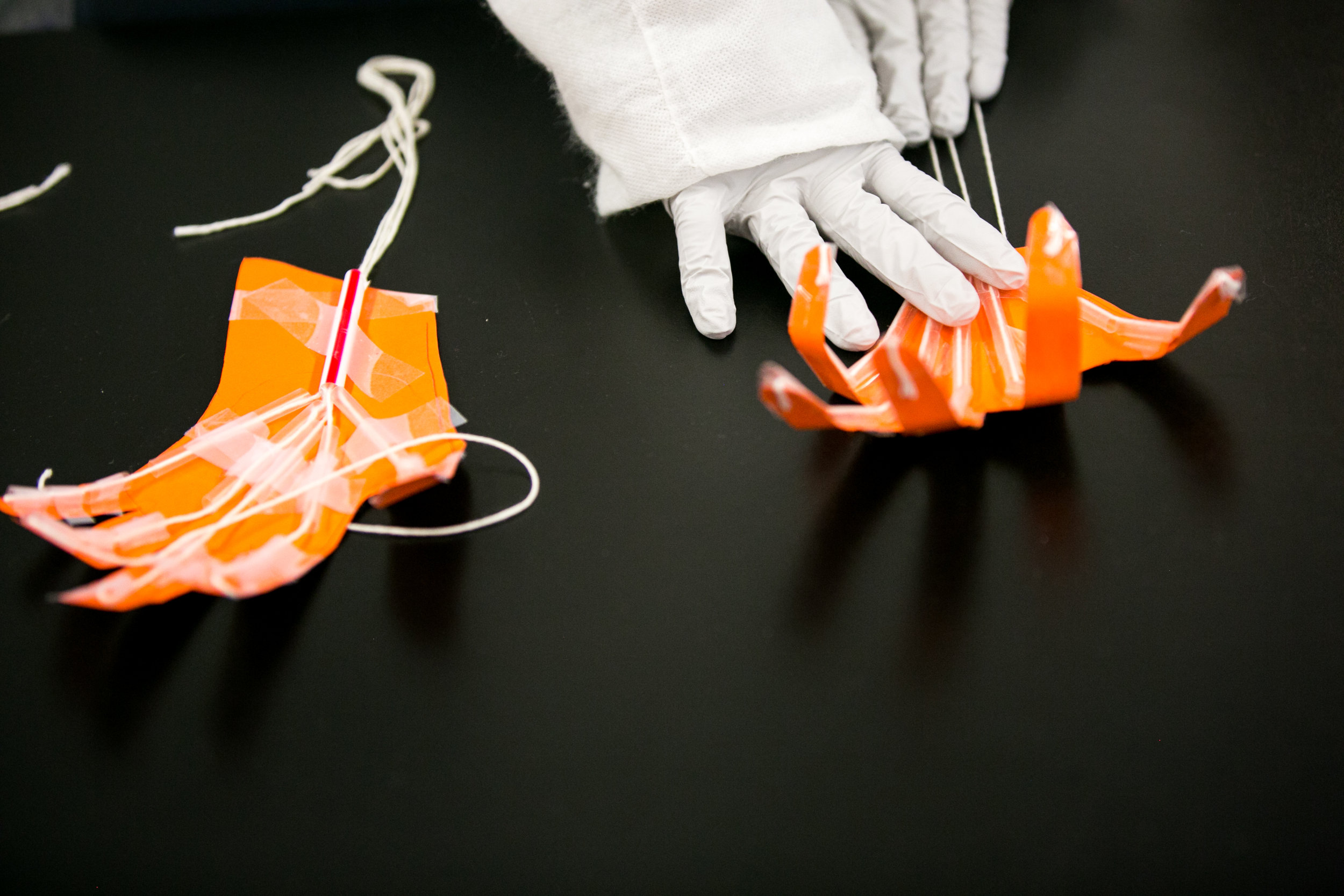
Testing the Model Hand

Testing the Model Hand

Testing the final model hand
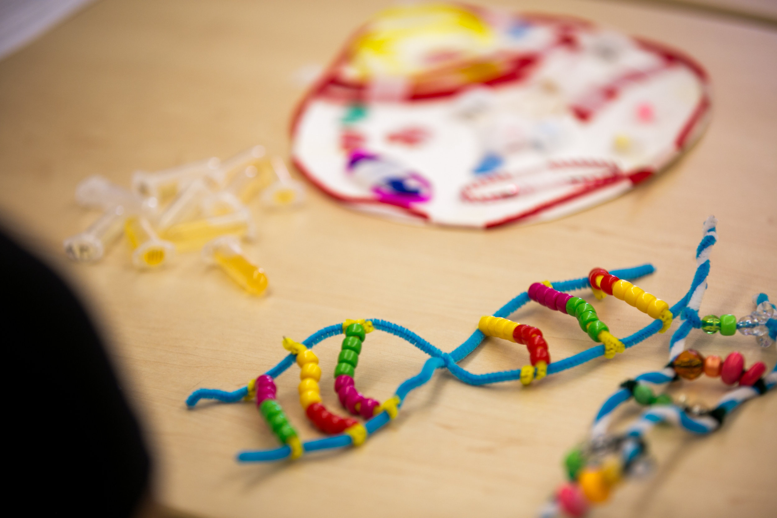
Completed DNA Model

Completed DNA Model

A finished DNA model with different colored beads representing each phosphate
20171027_KU_i2_STEM Week-ELIOT_022.jpg
Grybus-i2-Hurley-HR-4337.jpg
Grybus-i2-Perry-HR-1497.jpg
0088_i2_Sutton_088.JPG
0090_i2_Sutton_090.JPG
Cell Models

Cell Models

Student looks at a model of a cell

Student Looking into a Microscope

Student Looking into a Microscope

Student looks at a sample under the microscope
Grybus-i2-Perry-HR-1450.jpg
Adjusting the Magnification

Adjusting the Magnification

Student adjusts the magnification on the microscope

Microscope

Microscope

Student analyzes sample under the microscope

back of blood vessel.jpg
biopsy.jpg
blood vessel front.jpg
blood vessel with sickled cells.jpg
brain hat.jpg
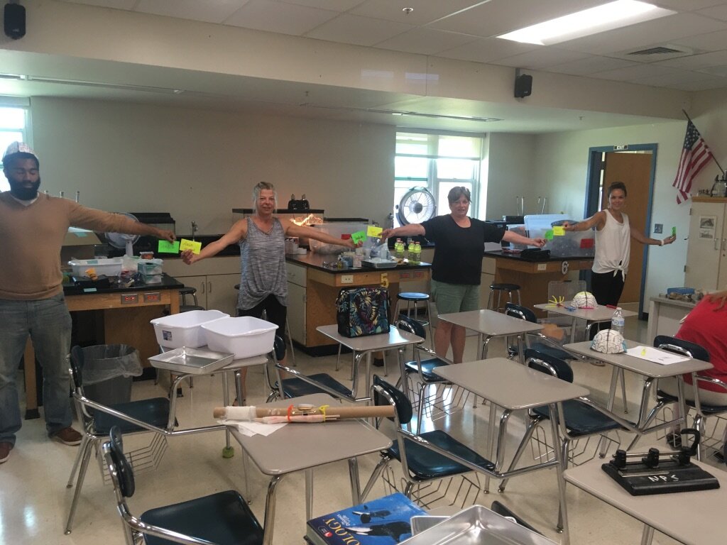
classroom neuron.jpg
classroom neurons.jpg
cutting into heart.jpg
heart.jpg
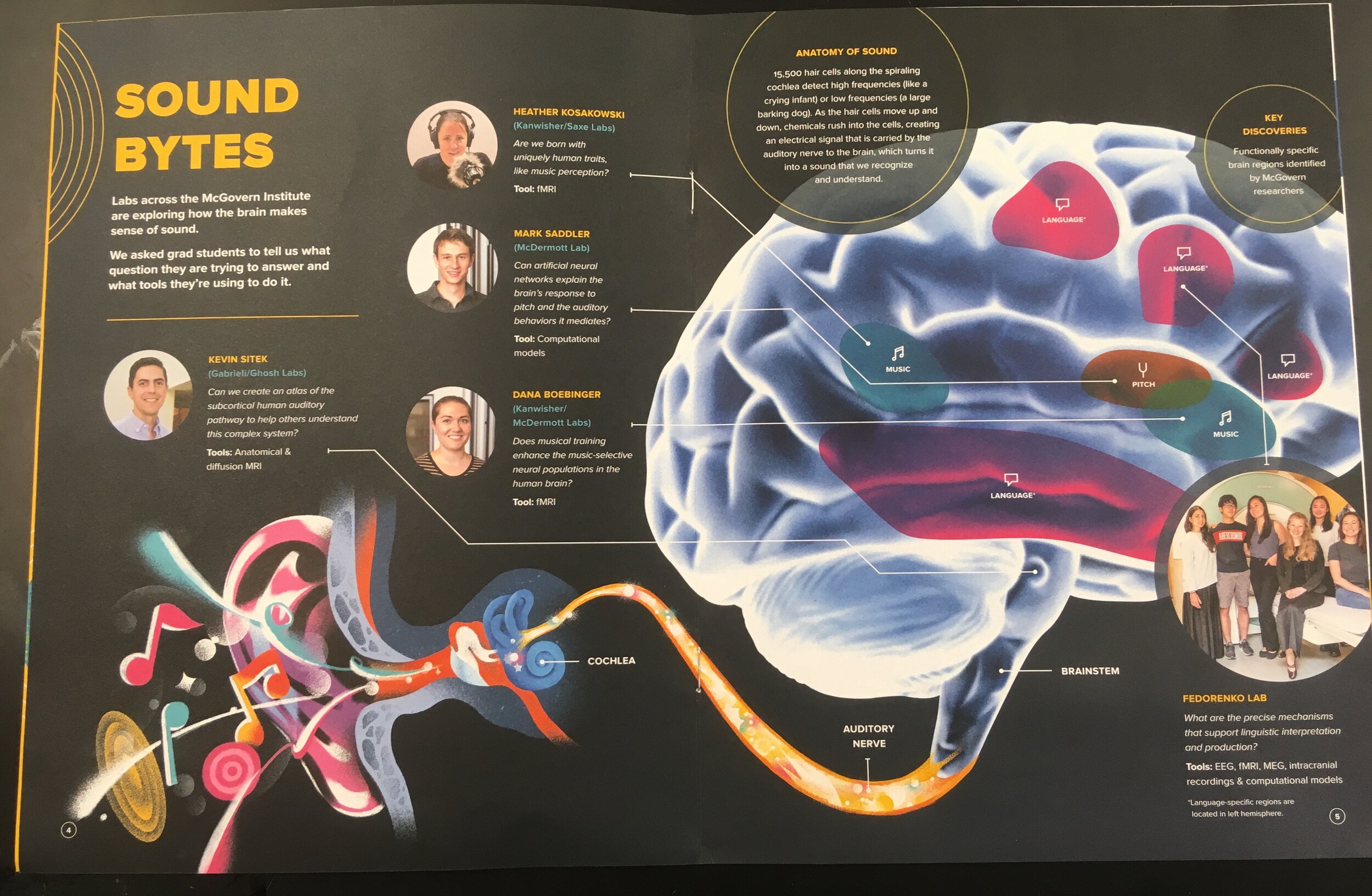
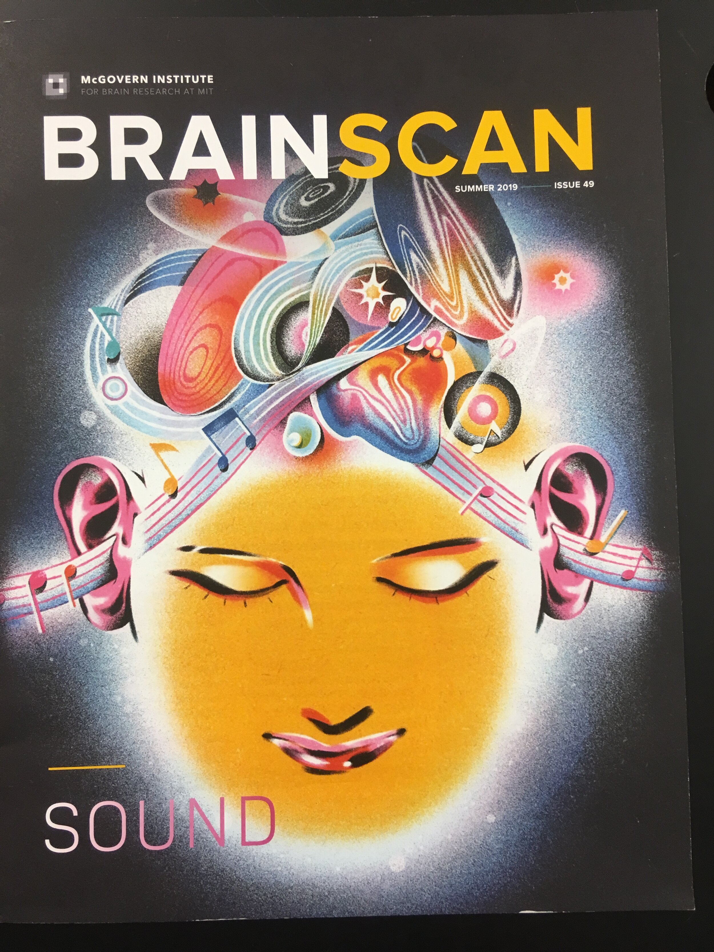
how sound affects the brain.jpg
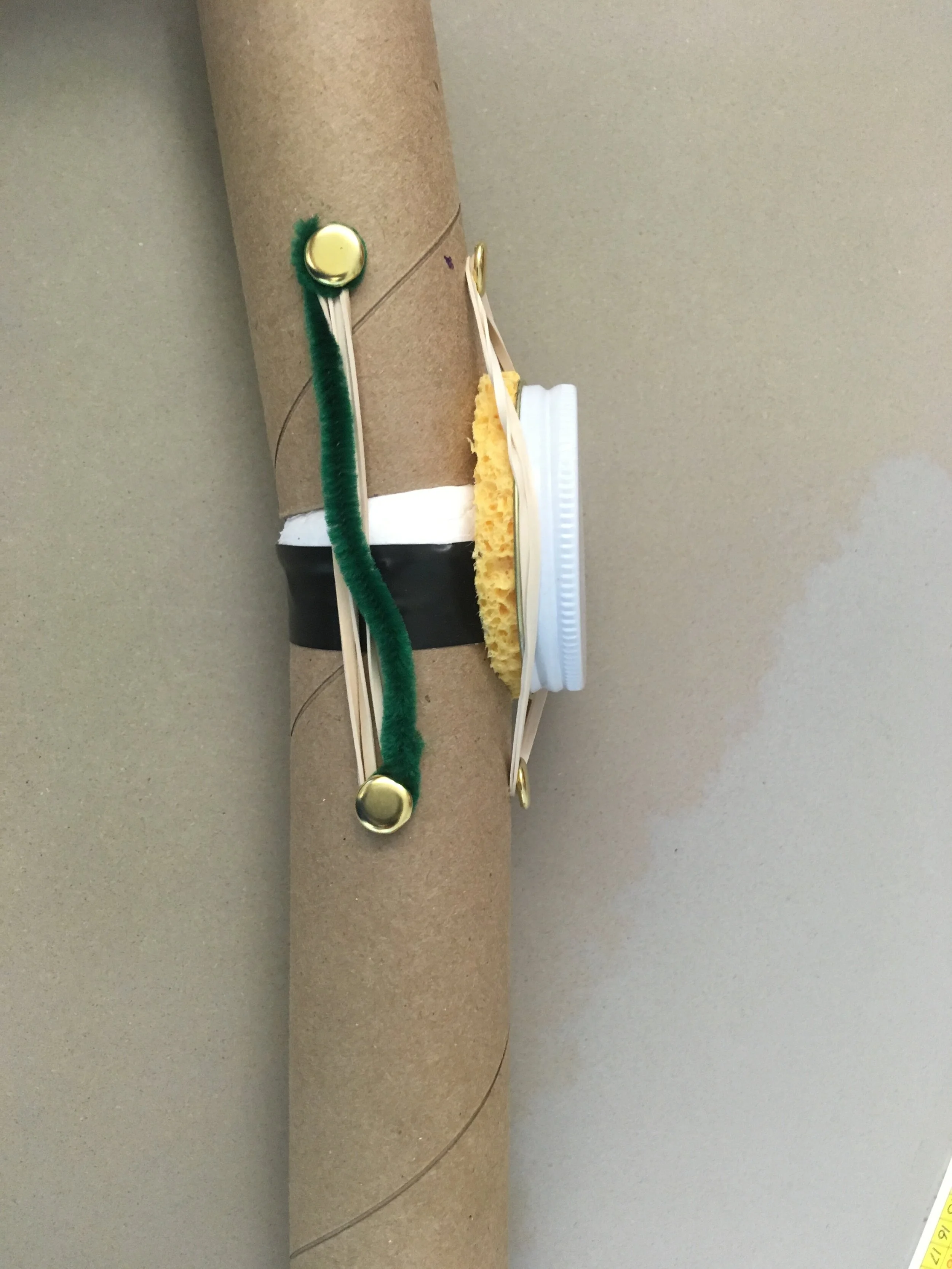
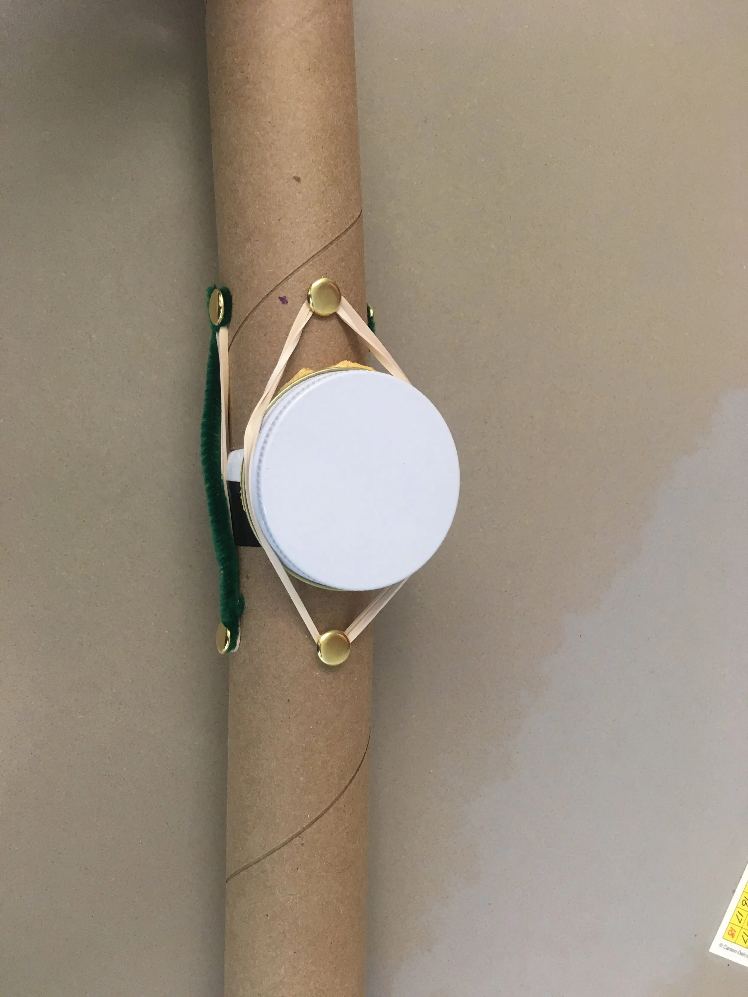
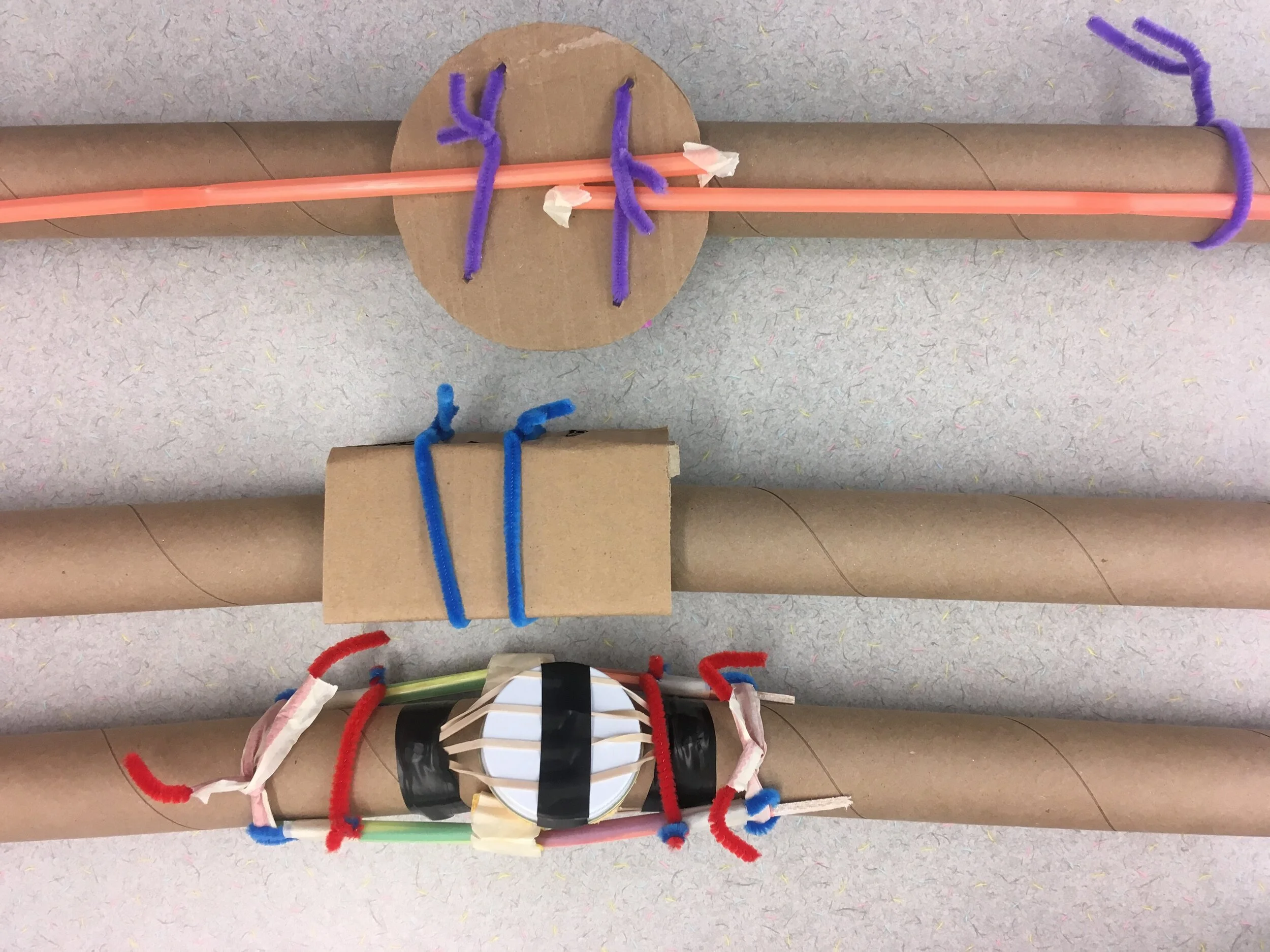
knee joint.JPG
knee joint(1).JPG
knee joint(2).JPG
kneecap.JPG
labeled heart with straws.jpg
patient_s leg in cast.JPG
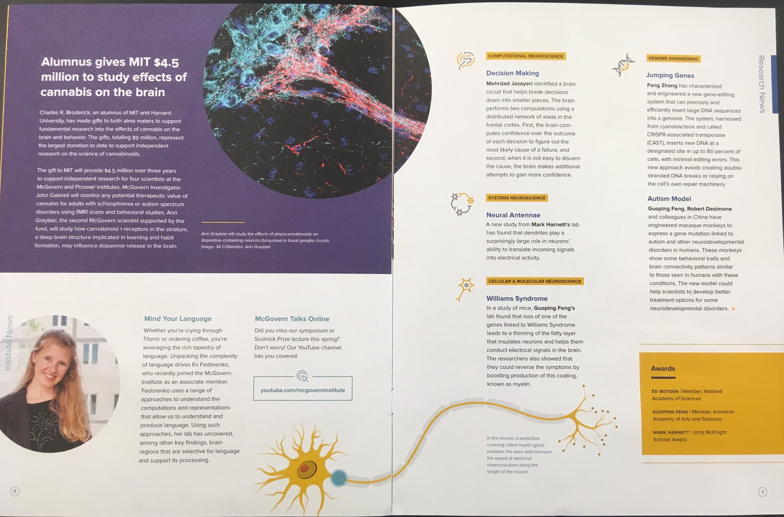
research brain MIT.JPG
research on brain MIT.jpg
research on brains at MIT.jpg
research on newborn brains.jpg
running stitch suture.jpg
running suture.jpg
setup for suture.jpg
Sickle cell in the news 2019 summer.jpg
sickled cells blocking blood vessel.jpg
sickled cells in blood vessel.jpg
simple interrupted suture.jpg
simple interrupted suture(1).jpg
splints by teachers.jpg
splints by teachers(1).jpg
splints by teachers(2).JPG
splints by teachers(3).JPG
1 2 3 4 5 6 7 8 9 10 11 12 13 14 15 16 17 18 19 20 21 22 23 24 25 26 27 28 29 30 31 32 33 34 35 36 37 38 39 40 41 42 43 44 45 46 47 48 49 50 51 52 53 54 55 56 57 58 59 60 61 62 63 64 65 66 67 68 69 70 71 72 73 74 75 76 77 78 79 80 81 82 83 84 85 86 87 88 89 90 91 92 93 94 95 96 97 98
Previous Next
Structure and Function of Blood: Video Tutorial
Heart Dissection
Heart Dissection
Heart Dissection
Heart Dissection
Heart Dissection
Heart Dissection
Heart with Blood Vessel Labels
Heart Dissection
Heart Dissection
Heart Dissection
Systems Working Together: Video Tutorial
Systems Working Together: Biopsy Simulation
Systems Working Together
DNA Isolation: Video Tutorial
Structure of a Finger: Video Tutorial
How to Suture: Video Tutorial
How to Suture
Model Neurons
Brain Hats
Brain Hats
Brain Hats
Brain Hats
Brain Hats
Brain Hats
Sheep Brain Dissection
Tools for the Sheep Brain Dissection
Sheep Brain Dissection
Model Patient
Model Patient
Model Patient
Model Patient
Model Patient
Model Patient
Model Patient
Showcase
Showcase
Showcase
Showcase
Showcase
Showcase
Showcase
Showcase
Showcase
Showcase
Showcase
Grybus-i2-Hurley-HR-4357.jpg
Creating the Model Hand
Testing the Model Hand
Completed DNA Model
20171027_KU_i2_STEM Week-ELIOT_022.jpg
Grybus-i2-Hurley-HR-4337.jpg
Grybus-i2-Perry-HR-1497.jpg
0088_i2_Sutton_088.JPG
0090_i2_Sutton_090.JPG
Cell Models
Student Looking into a Microscope
Grybus-i2-Perry-HR-1450.jpg
Adjusting the Magnification
Microscope
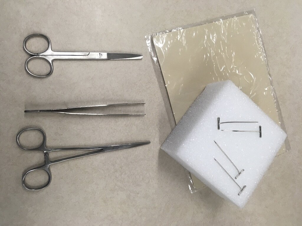
Biopsy Preparation
back of blood vessel.jpg
biopsy.jpg
blood vessel front.jpg
blood vessel with sickled cells.jpg
brain hat.jpg
classroom neuron.jpg
classroom neurons.jpg
cutting into heart.jpg
heart.jpg
how sound affects the brain.jpg
knee joint.JPG
knee joint(1).JPG
knee joint(2).JPG
kneecap.JPG
labeled heart with straws.jpg
patient_s leg in cast.JPG
research brain MIT.JPG
research on brain MIT.jpg
research on brains at MIT.jpg
research on newborn brains.jpg
running stitch suture.jpg
running suture.jpg
setup for suture.jpg
Sickle cell in the news 2019 summer.jpg
sickled cells blocking blood vessel.jpg
sickled cells in blood vessel.jpg
simple interrupted suture.jpg
simple interrupted suture(1).jpg
splints by teachers.jpg
splints by teachers(1).jpg
splints by teachers(2).JPG
splints by teachers(3).JPG
Back to Top
New i2 Logo - white.png
 

i2 Learning provides project-based STEM curriculum, professional development for teachers, and logistical support for educators to engage and inspire their students for a STEM-based world. i2 is a 501(c)3 organization which began in 2013 as a STEM summer program for 400 middle school children in Boston and New York and has since grown to reach almost 100,000 students in 48 states and 86 countries across the world.

Contact Us | Register